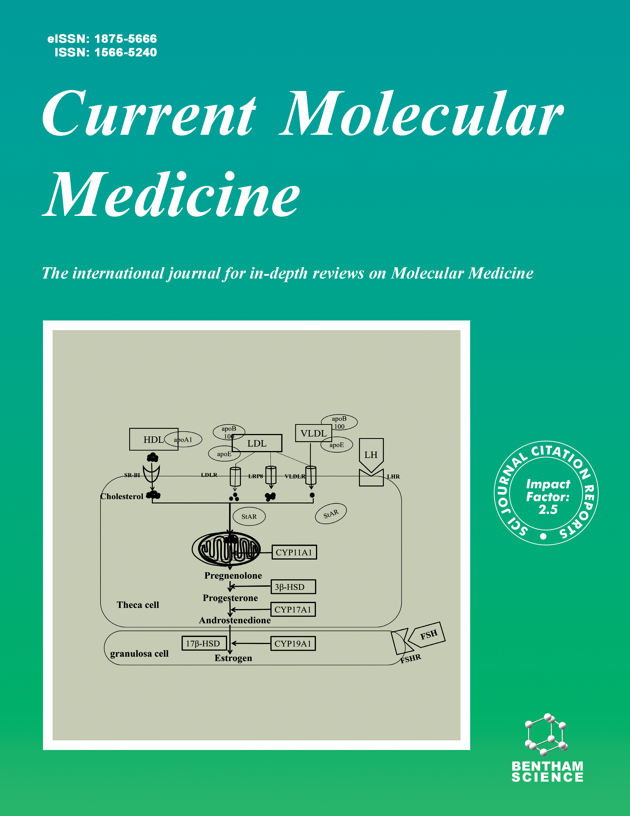
Current Molecular Medicine - Volume 25, Issue 5, 2025
Volume 25, Issue 5, 2025
-
-
Limning of HIF-2 and HIF-3 in the Tumor Microenvironment: Developing Concepts for the Treatment of Hypoxic Cancer
More LessAuthors: Suman Kumar Ray and Sukhes MukherjeeHypoxia, characterized by insufficient oxygen supply to tissues, is a significant factor in tumor growth and resistance to treatment. The hypoxia-inducible factor (HIF) signaling pathway is activated when oxygen levels decline, influencing cell activities and promoting tumor progression. HIF-1α and HIF-2α are the main targets for therapeutic intervention in tumors. Nevertheless, the significance of HIF-2α is often overlooked. This review examines the physiological role of HIF-2α in tumor growth and its involvement in tumor growth. HIFs, composed of hypoxia-responsive α and oxygen-insensitive β subunits, play a crucial role in controlling gene expression in both normal and solid tumor tissues under low oxygen levels. HIF-3α, formerly considered a detrimental modulator of HIF-regulated genes, exerts a transcriptional regulatory role by inhibiting gene expression through competition with HIF-1α and HIF-2α for binding to transcriptional sites in target genes under hypoxia. Recent research indicates that various HIF-3 variants exhibit distinct and potentially contrasting functionalities. Hypoxia often occurs during the initiation and progression of cancer formation. Recent research has discovered that HIF-2α, also known as endothelial PAS domain protein 1, has a significant impact on tumors. HIF-2α is a significant cancer-causing gene and a crucial predictor of prognosis in non-small cell lung cancer. However, due to limited research investigating the relationship between HIF-2α and small-cell lung cancer, it is not possible to reach a definitive conclusion. HIF-2α plays a vital function in cancer by preserving the stemness of cancer cells. This review provides a comprehensive overview of HIF-2 and the role of HIF-3 in various cancer-related processes, as well as its potential as a targeted therapeutic approach.
-
-
-
NLRP3 Inflammasome Triggers Inflammation of Obstructive Sleep Apnea
More LessAuthors: Xiaoting Yangzhong, Shu Hua, Yiqiong Wen, Xiaoqing Bi, Min Li, Yuanyuan Zheng and Shibo SunObstructive sleep apnea (OSA) is widespread in the population and affects as many as one billion people worldwide. OSA is associated with dysfunction of the brain system that controls breathing, which leads to intermittent hypoxia (IH), hypercapnia, and oxidative stress (OS). The number of NOD-like receptor family pyrin domain-containing (NLRP3) inflammasome was increased after IH, hypercapnia, and OS. NLRP3 inflammasome is closely related to inflammation. NLRP3 inflammasome causes a series of inflammatory diseases by activating IL-1β and IL-18. Subsequently, NLRP3 inflammasome plays an important role in the complications of OSA, including Type 2 diabetes (T2DM), coronary heart disease (CHD), hypertension, neuro-inflammation, and depression. This review will introduce the basic composition and structure of the NLRP3 inflammasome and focus on the relationship between the NLRP3 inflammasome and OSA and OSA complications. We can deeply understand how NLRP3 inflammasome is strongly associated with OSA and OSA complications.
-
-
-
Understanding the Roles of Non-coding RNAs and Exosomal Non-Coding RNAs in Diabetic Nephropathy
More LessAuthors: Yuye Zhu, Chunying Liu and Jamal HallajzadehOne of the greatest serious side effects of diabetes is diabetic nephropathy (DN), which is also the key factor in the sometimes-deadly diabetic end-stage renal disease. Progressive renal interstitial fibrosis is closely associated with oxidative stress, and the extracellular matrix is typically a feature of DN. Some RNAs formed by genome transcription that are not translated into proteins are recognized as non-coding RNAs. It has been shown that ncRNAs control apoptosis, inflammatory response, cell proliferation, autophagy, and other pathogenic processes, contributing to the pathogenesis of DN. Exosomes are nano-carriers vesicles that variety in size from 40 to 160 nm. Exosomes are widely present and dispersed in different bodily fluids, plentiful in nucleic acids, lipids, and proteins (microRNA, mRNA, tRNA, lncRNA, circRNA, etc.). Exosomes play a crucial role as messengers for cellular communication. They transport and transmit key signaling molecules, participate in the transfer of information and materials between cells, control cellular physiological processes, and are carefully linked to the beginning and development of many diseases. Herein, we summarized the role of different ncRNAs in DN. Moreover, we highlighted the role of the exosomal form of ncRNAs in the DN pathogenesis.
-
-
-
Ribosomal DNA and Neurological Disorders
More LessAuthors: Hong Zhou, Yuqing Xia, Rui Zhu, Yuemei Zhang, Xinming Zhang, Yongjian Zhang and Jun WangRibosomal DNA (rDNA) is important in the nucleolus and nuclear organization of human cells. Defective rDNA repeat maintenance has been reported to be closely associated with neurological disorders, such as Alzheimer’s disease, Huntington’s disease, Parkinson’s disease, amyotrophic lateral sclerosis, frontotemporal dementia, depression, suicide, etc. However, there has not been a comprehensive review on the role of rDNA in these disorders. In this review, we have summarized the role of rDNA in major neurological disorders to sort out the correlation between rDNA and neurological diseases and provided insights for therapy with rDNA as a target.
-
-
-
Targeting the Molecules in EMT: A Potential Therapeutic Opportunity in Breast Cancer
More LessAuthors: Siri Chandana Gampa and Sireesha V. GarimellaBreast Cancer (BC) is one of the most frequently occuring diseases in women, accounting for 90% of cancer-related deaths in women. Tumor cells can invade nearby tissues and spread to distant organs by metastasis. The epithelial-mesenchymal transition or EMT, which involves a number of transcription factors and signaling pathways, is a mechanism by which cells of the epithelium change into mesenchymal type capable of motility, invasion, and metastasis. EMT has grown to be a more intriguing target for developing cutting-edge treatment approaches since it is involved in diverse malignant transformation-related activities. Besides preventing tumor cell invasion and spread and the development of metastatic lesions, anti-EMT treatment methods also lessen cancer stemness and improve the efficacy of more traditional chemotherapeutics. EMT is, therefore, a desirable target in oncology. This review gives an overview of EMT, various markers of EMT, and different inhibitors used in therapies targeting EMT in BC.
-
-
-
A Systematic Review of the Impact of Resveratrol on Viral Hepatitis and Chronic Viral Hepatitis-related Hepatocellular Carcinoma
More LessBackgroundResveratrol (RSV) is used for the treatment of various diseases due to their anti-inflammatory and antioxidant activities. However, its beneficial aspects on viral hepatitis have been less investigated.
ObjectiveThis report reviews the impact of resveratrol on viral hepatitis and chronic viral hepatitis-related hepatocellular carcinoma (HCC).
MethodsThe systematic review was performed and reported according to the PRISMA 2020 statement. Several core databases, such as Cochrane Library, PubMed, Web of Science, EMBASE, and Scopus, were used for search on September 6, 2023. After extraction of the data, the desired information of the full text of the studies was recorded in Excel, and the outcomes and mechanisms were reviewed.
ResultsRSV inhibits viral replication through anti-HCV NS3 helicase activity, maintains redox homeostasis via glutathione (GSH) synthesis, improves T and B cell activity, and suppresses miR-155 expression. It also enhances viral replication by enhancing hepatitis C virus (HCV) RNA transcription, activating sirtuin-1 (SIRT1), which can increase peroxisome proliferator-activated receptor (PPAR), and SIRT1 activates the HBV X protein (HBx). Moreover, RSV is responsible for hepatitis-related HCC proliferation via suppression of mammalian target of rapamycin (mTOR), SIRT1 up-regulation, inhibiting expression of HBx, and reducing expression of cyclin D1.
ConclusionDespite the promising properties of RSV in inhibiting hepatitis-related HCC cell proliferation, its antiviral effects in viral hepatitis are controversial. The anti-hepatitis behaviors of RSV are mainly dose-dependent, and in some studies, activating some hepatoprotective pathways increases the transcription and replication of chronic HBV and HCV. Therefore, healthcare providers should be aware of viral hepatitis before using RSV supplements.
-
-
-
CCN3/NOV Serum Levels in Non-alcoholic Fatty Liver Disease (NAFLD) Patients in Comparison with the Healthy Group and its Correlation with TNF-α and IL-6
More LessAuthors: Reza Afrisham, Ghazal Alasvand, Yasaman Jadidi, Vida Farrokhi, Nariman Moradi, Shaban Alizadeh and Reza FadaeiIntroductionAdipokine irregularity leads to inflammation, endothelial dysfunction, insulin resistance (IR), and Non-Alcoholic Fatty Liver Disease (NAFLD). Previous studies linked NOV/CCN3 to obesity, IR, and inflammation, but no research has explored the connection between CCN3 serum levels and NAFLD.
MethodsThis case-control study assessed CCN3, IL-6, adiponectin, and TNF-α serum levels in 80 NAFLD patients and 80 controls using ELISA kits. Biochemical parameters were measured with commercial kits and an auto analyzer.
ResultsNAFLD patients exhibited significantly higher CCN3 (2399.85 ± 744.53 vs. 1712.84 ± 478.19 ng/ml), TNF-α, and IL-6 levels, and lower adiponectin levels compared to controls (P<0.0001). In the NAFLD group, CCN3 showed positive correlations with FBG, insulin, HOMA-IR, and TNF-α. Binary logistic regression analysis revealed increased NAFLD risk in the adjusted model (OR [95% CI] = 1.220 [1.315-1.131]). A CCN3 cut-off value of 1898.0050 pg/mL differentiated NAFLD patients from controls with 78.8% sensitivity and 73.2% specificity.
ConclusionIt was found that elevated CCN3 serum levels directly correlate with NAFLD incidence and inflammation markers (IL-6 and TNF-α). CCN3 could serve as a potential biomarker for NAFLD, but further research is needed to validate this finding and assess its clinical utility.
-
-
-
Anti-Cancer and Anti-Oxidant Effects of Fenoferin-loaded Human Serum Albumin Nanoparticles Coated with Folic Acid-bound Chitosan
More LessBackgroundSeveral diseases, including cancer, can be effectively treated by altering the nanocarrier surfaces so that they are more likely to be targeted.
ObjectiveThis study aimed to prepare human albumin (HSA) nanoparticles containing Fenoferin (FN) modified with folic acid (FA) attached to Chitosan (CS) to improve its anti-cancer properties.
MethodsNanoparticles were first synthesized and surface modified. Their physicochemical properties were assessed by different methods, such as FESEM, FTIR, and DLS. In addition, the percentage of drug encapsulated was measured by indirect method. Besides evaluating the cytotoxic effects of nanoparticles using the MTT assay, the antioxidant capacity of FN-HSA-CS-FA was assessed using the ABTS and DPPH methods. Nanoparticles were also investigated for their anti-cancer effects by evaluating the expression of apoptosis and metastasis genes.
ResultsBased on this study, FN-HSA-CS-FA was 165.46 nm in size, and a uniform dispersion distribution was identified. Particles were reported to have a zeta potential of +29 mV, which is within the range of stable nanoparticles. Approximately 75% of FN is encapsulated in nanoparticles. Cytotoxic assay determined that liver cancer cells were most sensitive to treatment with an IC50 of 144 µg/ml. Inhibition of free radicals by nanoparticles is estimated to have an IC50 value of 195.23 and 964 µg/ml, for ABTS and DPPH, respectively. In the treatment with nanoparticles, flow cytometry results of arresting the cells in the SubG1 phase and real-time qPCR results indicated increased expression of caspases-3, caspase-8, and caspase-9 genes.
ConclusionAccording to this study, synthesized nanoparticles inhibited free radicals and activated apoptosis in liver cancer cells, and the capability of these nanoparticles to inhibit cancer cells was also confirmed. This formulation can, therefore, be used in preclinical studies to test the efficacy of the drug.
-
-
-
CXCL13-neutralizing Antibody Alleviate Chronic Skeletal Muscle Degeneration in a Mouse Model
More LessAuthors: Zhongcheng Xie, Jimin Yang, Chunmeng Jiao, Hui Chen, Siyu Ouyang, Zhiyang Liu, Qin Hou and Jifeng LiuIntroductionSkeletal muscle degeneration is a common effect of chronic muscle injuries, including fibrosis and fatty infiltration, which is the replacement of pre-existing parenchymal tissue by extracellular matrix proteins and abnormal invasive growth of fibroblasts and adipocytes.
MethodsThis remodeling limits muscle function and strength, eventually leading to reduced quality of life for those affected. Chemokines play a major role in the regulation of immunocyte migration, inflammation, and tissue remodeling and are implicated in various fibrotic and degenerative diseases. In this study, we aimed to investigate the role of the B-cell chemokine CXCL13 in the gastrocnemius muscle of the Achilles tendon rupture model mouse. We hypothesize that CXCL13 may promote fibrosis and aggravate skeletal muscle degeneration. We performed RNA sequencing and bioinformatics analysis of gastrocnemius muscle from normal and model mice to identify differentially expressed genes and signal pathways related to skeletal muscle degeneration and fibrosis.
ResultsOur results show that CXCL13 is highly expressed in chronically degenerating skeletal muscle. Furthermore, CXCL13-neutralising antibodies with therapeutic potential were observed to inhibit fibrosis and adipogenesis in vivo and in vitro.
ConclusionOur study reveals the underlying therapeutic implications of CXCL13 inhibition for clinical intervention in skeletal muscle degeneration, thereby improving patient prognosis.
-
-
-
Establishment and Validation of Lactate Metabolism-Related Genes as a Prognostic Model for Gastric Cancer
More LessAuthors: Jinyu Hu, Qinxuan Xu, Yuchang Fei, Zhengwei Tan and Lei PanBackgroundGastric Cancer (GC) has become one of the most important causes of cancer-related deaths worldwide due to its intractability. Studying the mechanisms of gastric carcinogenesis, recurrence, and metastasis, and searching for new therapeutic targets have become the main directions of today's gastric cancer research. Lactate is considered a metabolic by-product of tumor aerobic glycolysis, which can regulate tumor development through various mechanisms, including cell cycle regulation, immunosuppression, and energy metabolism. However, the effects of genes related to lactate metabolism on the prognosis and tumor microenvironmental characteristics of GC patients are unknown.
MethodsIn this study, we have collected gene expression data of gastric cancer from The Cancer Genome Atlas (TCGA) and identified differentially expressed genes in gastric cancer using the “Limma” software package.
Results76 differentially expressed lactate metabolism-related genes were screened, and then the Least Absolute Shrinkage and Selection Operator (LASSO) and Cox regression analysis were employed that identified 8 genes, constructed Lactate Metabolism-related gene signals (LMRs), and verified the reliability of the prognostic risk mapping by using TCGA training set and TCGA internal test set. Finally, the functional enrichment analysis was employed to identify the molecular mechanism.
ConclusionEight lactate metabolism-related genes were constructed into a new predictive signal that better predicted the overall survival of gastric cancer patients and can guide clinical decisions for more precise and personalized treatment.
-
-
-
Evolutionary Sequences and Structural Information-driven Reconstruction of New Insulin-like Growth Factor-I Peptide Variants
More LessAuthors: Nazam Khan, Maryam Althobiti, Raj Kumar Chinnadurai, Samir Alharbi and Rajender KumarBackgroundInsulin-like growth factor-I (IGF-I) is crucial in controlling cell growth, proliferation, and apoptosis. Its strong link to the development of cancers such as breast, prostate, lung, thyroid, and colorectal has positioned the IGF-1 signalling pathway as a promising target for novel cancer therapies. When activated, the IGF-1 receptor (IGF-1R) binds to IGF-I, playing a central role in promoting tumour cell growth and survival.
MethodsIn this study, we combined evolutionary sequences with structural and functional data of IGF-1 to reconstruct ancestral sequences and design novel IGF-1 peptide variants.
ResultsThe insulin-like growth factor system exhibits a vast sequence diversity, yet it shares a similar structural topology with conserved three pairs of disulfide linkages. Our study reveals that IGF-1 is associated with the IGF system of cell surface receptors through protein-protein interactions. Reconstructed IGF-1 variants show similar structure fold to reported viral IGF-1 competitive antagonists.
ConclusionThis new insight guides the design of novel natural IGF-1 mimic peptides. It enhances our understanding of IGF-1's functionality and opens new avenues for the development of therapeutic peptides and small molecules as anti-cancer agents.
-
Volumes & issues
-
Volume 25 (2025)
-
Volume 24 (2024)
-
Volume 23 (2023)
-
Volume 22 (2022)
-
Volume 21 (2021)
-
Volume 20 (2020)
-
Volume 19 (2019)
-
Volume 18 (2018)
-
Volume 17 (2017)
-
Volume 16 (2016)
-
Volume 15 (2015)
-
Volume 14 (2014)
-
Volume 13 (2013)
-
Volume 12 (2012)
-
Volume 11 (2011)
-
Volume 10 (2010)
-
Volume 9 (2009)
-
Volume 8 (2008)
-
Volume 7 (2007)
-
Volume 6 (2006)
-
Volume 5 (2005)
-
Volume 4 (2004)
-
Volume 3 (2003)
-
Volume 2 (2002)
-
Volume 1 (2001)
Most Read This Month


