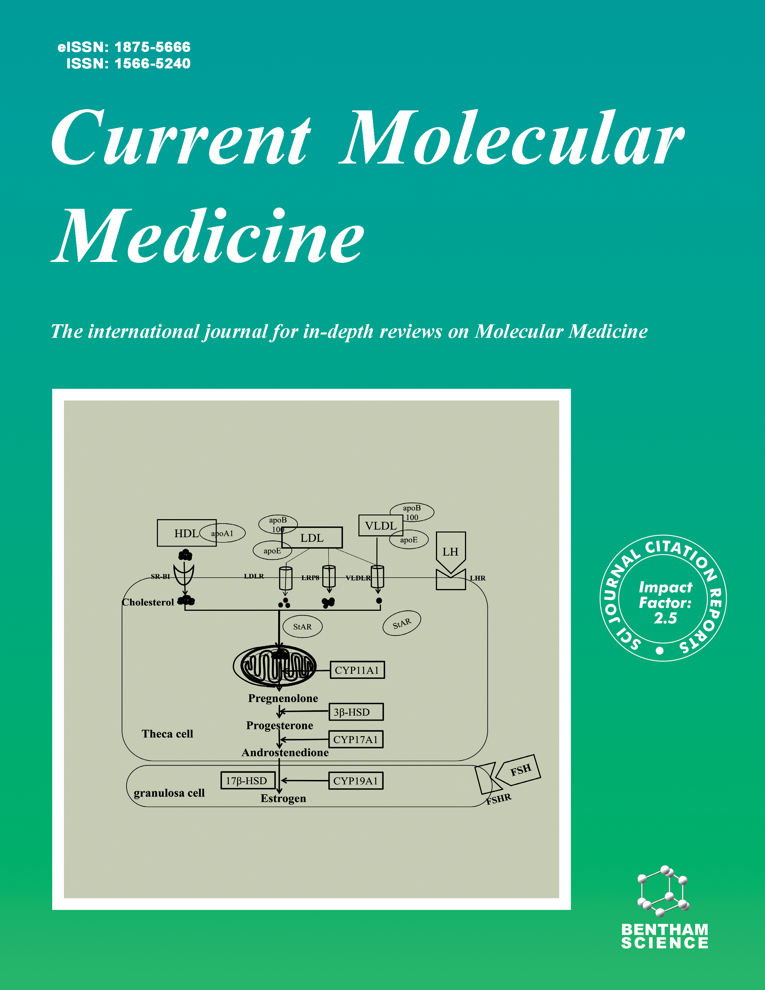
Current Molecular Medicine - Volume 14, Issue 9, 2014
Volume 14, Issue 9, 2014
-
-
Reactive Oxygen Species: Physiological Roles in the Regulation of Vascular Cells
More LessReactive oxygen species (ROS) are now appreciated to play several important roles in a number of biological processes and regulate cell physiology and function. ROS are a heterogeneous chemical class that includes radicals, such as superoxide ion (O2•), hydroxyl radical (OH•) and nitric oxide (NO•), and non-radicals, such as hydrogen peroxide (H2O2), singlet oxygen (1O2), hypochlorous acid (HOCl), and peroxynitrite (NO3 -). In the cardiovascular system, besides playing a critical role in the development and progression of vasculopathies and other important pathologies such as congestive heart failure, atherosclerosis and thrombosis, ROS also regulate physiological processes. Evidence from a wealth of cardiovascular research studies suggests that ROS act as second messengers and play an essential role in vascular homeostasis by influencing discrete signal transduction pathways in various systems and cell types. They are produced throughout the vascular system, regulate differentiation and contractility of vascular smooth muscle cells, control vascular endothelial cell proliferation and migration, mediate platelet activation and haemostasis, and significantly contribute to the immune response. Our understanding of ROS chemistry and cell biology has evolved to the point of realizing that different ROS have distinct and important roles in cardiovascular physiology. This review will outline sources, functions and molecular mechanisms of action of different ROS in the cardiovascular system and will describe their emerging role in healthy cardiovascular physiology and homeostasis.
-
-
-
Cholinergic Receptors as Target for Cancer Therapy in a Systems Medicine Perspective
More LessAuthors: P. Russo, A. Del Bufalo, M. Milic, G. Salinaro, M. Fini and A. CesarioEpithelial cells not innervated by cholinergic neurons express nicotinic and muscarinic acetylcholine (ACh) receptors (nAChR, mAChR). nAChR and mAChR are components of the auto-/paracrine-regulatory loop of non-neuronal ACh release. The cholinergic control of non-neuronal cells may be mediated by different effects (synergistic, additive, or reciprocal) triggered by these receptors. The ionic events (Ca+2 influx) are generated by the ACh-opening of nAChR channels, while the metabolic events by ACh-binding to G-proteincoupled mAChR. Effective inter- and intracellular signaling is crucial for valuable cancer cells proliferation and survival. Depending on cancer cell type, different AChR have been identified. The proliferation of airways epithelial cancer cells and pancreatic cancer cells may be under the control of α7-nAChR and M3-mAChR, while breast cancer cells and colon cancer cells are regulated by α9-nAChR, and M3-mAChR, respectively. In turn, these receptors may activate different pathways (Ras-Raf-1-Erk-AKT) as well as other receptors (β- adrenergicR). nAChR or mAChR antagonists may inhibit cancer growth. Inhibition of M3 by antisense or antagonists (Darifenacin, Tiotropium) reduces lung or colon cancer proliferation, as well as inhibition of α9- nAChR [polyphenol (-)-epigallocatechin-3-gallate] diminishes breast cancer cells growth. α7-nAChR silencing inhibits lung cancer proliferation. Moreover, inhibition of the nAChR-β-adrenergicR pathway (β-blockers) could be also useful. This review will describe the future translational perspectives of cholinergic receptors druginhibition in a complex disease such as cancer that poses compelling treatment challenges. Cancer happens as consequence of disease-perturbed molecular networks in relevant organ cells that change during progression. The framework for approaching these challenges is a systems approach.
-
-
-
Recent Studies on the Antimicrobial Peptides Lactoferricin and Lactoferrampin
More LessLactoferricin and lactoferrampin, peptides derived from the whey protein lactoferrin, are antimicrobial agents with a promising prospect and are currently one of the research focuses. In this review, a basic introduction including location and solution structures of these two peptides is given. Their biological activities encompassing antiviral, antibacterial, antifungal and anti-inflammatory activities with possible mechanisms are mentioned. In terms of modification studies, research about identification of their active derivatives and crucial amino acid residues is also discussed. Various attempts at modification of lactoferricin and lactoferrampin such as introducing big hydrophobic side-chains; employing special amino acids for synthesis; N-acetylization, amidation, cyclization and peptide chimera are summarized. The studies on lactoferricin-lactoferrampin chimera are discussed in detail. Future prospects of lactoferricin and lactoferrampin are covered.
-
-
-
Human Cognitive and Neuro-Psychiatric Bio-Markers in the Cardiac Peri-Operative Patient
More LessSome of the complexities of surgical interventions include neurological and psychiatric disturbances. Prompt identification and early treatment of these complications are pivotal in achieving excellent clinical results. Recognizing major adverse events such as stroke, seizure or delirium is usually straight-forward, however the discovery of less frequent or more subtle post-operative changes such as cognitive dysfunction might be delayed due to lack of appropriate diagnostic tools. This review summarizes biological markers that can be utilized as surrogates in evaluating surgery-related neuro-psychiatric disorders.
-
-
-
Prolylcarboxypeptidase Independently Activates Plasma Prekallikrein (Fletcher Factor)
More LessAuthors: J. Wang, A. Matafonov, H. Madkhali, F. Mahdi, D. Watson, A.H. Schmaier, D. Gailani and Z. Shariat-MadarProlylcarboxypeptidase isoform 1 (PRCP1) is capable of regulating numerous autocrines and hormones, such as angiotensin II, angiotensin III, αMSH1-13, and DesArg9 bradykinin. It does so by cleaving a C-terminal PRO-X bond. Recent work also indicates that the human PRCP1 activates plasma prekallikrein (PK) to kallikrein on endothelial cells through an uncharacterized mechanism. This study aims to identify PRCP1 binding interaction and cleavage site on PK. Recently, a cDNA encoding a novel splice variant of the human PRCP1 was identified. This isoform differed only in the N-terminal region of the deduced amino acid sequence. Using structural and functional studies, a combination of peptide mapping and site-directed mutagenesis approaches were employed to investigate the interaction of PRCP1 with PK. Three PRCP peptides, in decreasing order of potency, from 1) the N-terminus of the secreted protein, 2) spanning the opening of the active site pocket, and 3) in the dimerization region inhibit PRCP activation of PK on endothelial cells. Investigations also tested the hypothesis that PRCP cleavage site on PK is between its C-terminal Pro 637 (P637) and Ala 638 (A638). Recombinant forms of PK with C-terminal alanine mutagenesis or a stop codon is activated equally as wild type PK by PRCP. In conclusion, PRCP1 interacts with PK at multiple sites for PK activation. PRCP1 also enhances FXIIa activation of PK, suggesting that its activation site on PK is not identical to that of FXIIa.
-
-
-
Ipsilateral Hippocampal Proteomics Reveals Mitochondrial Antioxidative Stress Impairment in Cortical-Lesioned Chronic Mild Stressed Rats
More LessIn this study, a two-dimensional gel-based proteomic approach was applied to profile the protein alterations underlying the significant adverse effects from post-stroke depression (PSD). In view of the close association between left prefrontal cortical dysfunction and PSD, a PSD rat model was constructed through a left anterior cortical lesion followed by chronic mild stress (CMS) for three weeks. Through sucrose preference testing, PSD rats displayed depression-like behavior during the entire CMS period. In contrast, stroke rats displayed depression-like behavior in the first week post-stroke and recovered in the second week post-stroke. To investigate the PSD-induced protein expression changes, ipsilateral hippocampal protein expression in stroke, PSD, and control rats were comparatively analyzed. 46 differential proteins were identified, 22 of which were regulated in opposing directions by stroke and post-stroke stress. The majority of these 22 proteins were involved in neurogenesis, cytoskeletal remodeling, and energy metabolism. Additional proteins were functionally related to mitochondrial antioxidative stress systems. The differential proteins expressed in opposing directions by stroke and post-stroke stress may play a role in self-repair after adult brain lesions, suggesting that stroke induces self-repair mechanisms, while post-stoke stress mitigates them, in the rat hippocampus. Among these differential proteins dysregulated in opposing directions, three mitochondrial proteins involved in mitochondrial antioxidative stress – heat shock 70 kDa protein 9, peroxiredoxin-6, and prohibitin – were validated and may play an important role in stroke-injury self-repair and PSD-induced injury of hippocampal neurons. These findings offer new insight into deciphering the molecular mechanisms underpinning PSD's adverse effects on stroke recovery.
-
-
-
The Tumor Suppressor, p53 Regulates the γA-Crystallin Gene During Mouse Lens Development
More LessAuthors: X.-H. Hu, Q. Nie, M. Yi, T.-T. Li, Z.-F. Wang, Z.-X. Huang, X.-D. Gong, L. Zhou, W.-K. Ji, W.-F. Hu, J.-F. Liu, L. Wang, Z. Woodward, J. Zhu, W.-B. Liu, Q.D. Nguyen and D.W.-C. LiThe tumor suppressor, p53 regulates a large number of target genes to control cell proliferation and apoptosis. In addition, it is also implicated in the regulation of cell differentiation in muscle, the circulatory system and various carcinoma tissues. We have recently shown that p53 also controls lens differentiation. Regarding the mechanism, we reveal that p53 directly regulates several genes including c-Maf and Prox1, two important transcription factors for lens differentiation, and αA and βA3/A1, the lens differentiation markers. In the present study, we present evidence to show that the γA-crystallin gene distal promoter and the first intron also contain p53 binding sites and are capable of mediating p53 control during mouse lens development. First, gel mobility shifting assays revealed that the p53 protein in nuclear extracts from human lens epithelial cells (HLE) directly binds to the p53 binding sites present in the γA-crystallin gene. Second, the exogenous wild type p53 induces the dose-dependent expression of the luciferase reporter gene driven by the basic promoter containing the γA-crystallin gene p53 binding site. In contrast, the exogenous dominant negative mutant p53 causes a dose-dependent inhibition of the same promoter. Third, ChIP assays revealed that p53 binds to the γA-crystallin gene promoter in vivo. Finally, in the p53 knockout mouse lenses, the expression level of the γAcrystallin gene was found attenuated in comparison with that in the wild type mouse lenses. Together, our results reveal that p53 regulates γA-crystallin gene expression during mouse lens development. Thus, p53 directly regulates all 3 types of crystallin genes to control lens differentiation.
-
-
-
Evidence for Up-Regulation of Purinergic Receptor Genes Associating with TRPV1 Receptors and Neurotrophic Factors in the Inflamed Human Esophagus
More LessAuthors: K.-R. Shieh, S.-C. Yang, H.-L. Tseng, C.-H. Yi, T.-T. Liu and C.-L. ChenPurinergic receptors are implicated in nociceptive signaling in small primary afferents via activation of adenosine triphosphate (ATP). ATP appears to mediate HCl-induced transient receptor potential vanilloid receptor 1 (TRPV1) activation in esophageal mucosa. Up-regulation of TRPV1 expression in gastroesophageal reflux disease (GERD) is associated with increased nerve growth factor (NGF) and glial derived neurotrophic factor (GDNF). This study aims to genetically determine the expression of purinergic receptors in severe inflamed human esophagus. Distal esophageal biopsies from the subjects with erosive GERD, asymptomatic patients (AP) and healthy ones were examined. Using real-time qPCR for detecting purinergic receptors (P2X2, P2X3, P2X7, P2Y1, P2Y2, P2Y4, P2Y6 and P2Y12), TRPV1, TRPV4, NGF, and GDNF was done in this study. Both P2X3 and P2X7 mRNA expressions in GERD patients significantly increased than those in healthy controls (P < 0.001) and AP (P < 0.001), but P2X2, P2Y1, P2Y2, P2Y4, P2Y6, P2Y12 or P2Y12 had no difference within the control, AP or GERD subjects. The well correlated expression in P2X3 gene with TRPV1 (r = 0.46, P = 0.002), NGF (r = 0.54, P = 0.0002), and GDNF (r = 0.64, P = 0.0001) was found. The P2X7 gene expressions also well correlated with TRPV1 (r = 0.47, P = 0.002), NGF (r = 0.32, P = 0.037), and GDNF (r = 0.42, P = 0.005). These results suggest that chronic esophagitis increases mRNA expressions of P2X3 and P2X7 receptors accompanied by up-regulation of TRPV1 and neurotrophic factors (NGF and GDNF). These genetical alterations in esophageal mucosa might mediate sensitization of inflamed human esophagus.
-
-
-
A New Approach of Delivering siRNA to the Cornea and its Application for Inhibiting Herpes Simplex Keratitis
More LessSmall interfering RNA (siRNA) is a potential agent for the treatment of ocular surface diseases. Previous studies delivered siRNA by directly injecting siRNA into cornea or conjunctiva. In the present study we sought to explore an alternative approach to deliver siRNA into mouse cornea via eye drops that contains cy3-labeled siRNA (cy3-siRNA) and different cationic complexing agents and to evaluate the effects of siRNA targeting HSV-1 ICP4 gene (ICP4-siRNA) on mouse herpes simplex keratitis (HSK). Cy3-siRNA was mixed with Lipofectamine 2000, EntransterTM-in vivo, polyethyleneimine (PEI) or PEO-PPO-PEO polymers at different ratios. The efficacy of delivery was analyzed after topical application of the complexes to normal, EDTA treated, and epithelial scraped cornea of BALB/c mouse eyes. Compared to the other delivery agents and schedules, PEI at 0.75 mg/ml with 20 μM cy3-siRNA complex delivered eight times daily for two days was the most efficient as revealed by its production of the greatest fluorescence in cornea epithelial cells. In mouse HSK, the application of ICP4-siRNA+PEI eye drops reduced the damage to the corneal epithelia and decreased viral VP16 expression in the corneal tissue. These results proved the idea that siRNA can be formulated into eye drops with carriers for effective delivery into the cornea and that the formulated eye drops containing ICP4-siRNA can inhibit HSV-1 replication in the mouse corneas.
-
-
-
The Effect of Claudin-5 Overexpression on the Interactions of Claudin-1 and -2 and Barrier Function in Retinal Cells
More LessClaudin-5, one of the dominant tight junctions (TJs) proteins, plays an important role in maintaining the barrier function in the blood brain and retinal barrier. This study aimed to investigate the effect of claudin-5 overexpression on the interactions of claudin-1 and -2 and barrier functions in primary cultured human retinal pigment epithelium cells (HRPECs) and human retina endothelial cells (HRECs). Lentivirus was used to mediate the overexpression of claudin-5 in retinal cells. Significantly increased mRNA and protein levels of claudin-5 were detected in the transfection group. After the transfected cells grew on the transwell membrane for three weeks, a stable monolayer cell barrier model was established in vitro. The claudins expressions analysis showed that overexpressed claudin-5 significantly increased the expression of claudin-1, while it decreased the expression of claudin-2 in both mRNA and protein level. Co-IP experiments and barrier function assay revealed that claudin-5 overexpression promoted the interactions of claudin-1 and claudin-2 and enhanced the barrier function of retinal cells. Intriguingly, the exogenous expression of claudin-5 induced new interaction pattern between claudin-5 and claudin-1 or -2 in HRPECs, which do not have endogenous claudin-5 expression. In addition, claudin-5 overexpression decreased cell mobility and the sprouting capability of vessel tube formation in vitro. This study demonstrated that claudin-5 has a positive regulation in the formation of retinal barrier. Claudin elements and their interactions can be modulated and that such dynamic properties are important for the functions of TJs, ranging from the regulation of retinal barrier integrity to junction-associated signaling mechanisms.
-
-
-
The Yin and Yang of Inflammation
More LessAuthors: M.A. Blackman, J.L. Yates, C.M. Spencer, E.E. Vomhof-DeKrey, A.M. Cooper and E.A. LeadbetterInflammation is an essential protective part of the body’s response to infection, yet many diseases are the product of inflammation. For example, inflammation can lead to autoimmune disease and tissue damage, and is a key element in chronic health conditions such as heart disease, diabetes, rheumatoid arthritis, and also drives changes associated with aging. Animal models of infectious and chronic disease are important tools with which to dissect the pathways whereby inflammatory responses are initiated and controlled. Animal models therefore provide a prism through which the role of inflammation in health and disease can be viewed, and are important means by which to dissect mechanisms and identify potential therapies to be tested in the clinic. A meeting, “The Yin and Yang of Inflammation” was organized by Trudeau Institute and was held between April 4-6, 2014. The main goal was to bring together experts from biotechnology and academic organizations to examine and describe critical pathways in inflammation and place these pathways within the context of human disease. A group of ~80 scientists met for three days of intense formal and informal exchanges. A key focus was to stimulate interactions between basic research and industry.
-
Volumes & issues
-
Volume 25 (2025)
-
Volume 24 (2024)
-
Volume 23 (2023)
-
Volume 22 (2022)
-
Volume 21 (2021)
-
Volume 20 (2020)
-
Volume 19 (2019)
-
Volume 18 (2018)
-
Volume 17 (2017)
-
Volume 16 (2016)
-
Volume 15 (2015)
-
Volume 14 (2014)
-
Volume 13 (2013)
-
Volume 12 (2012)
-
Volume 11 (2011)
-
Volume 10 (2010)
-
Volume 9 (2009)
-
Volume 8 (2008)
-
Volume 7 (2007)
-
Volume 6 (2006)
-
Volume 5 (2005)
-
Volume 4 (2004)
-
Volume 3 (2003)
-
Volume 2 (2002)
-
Volume 1 (2001)
Most Read This Month


