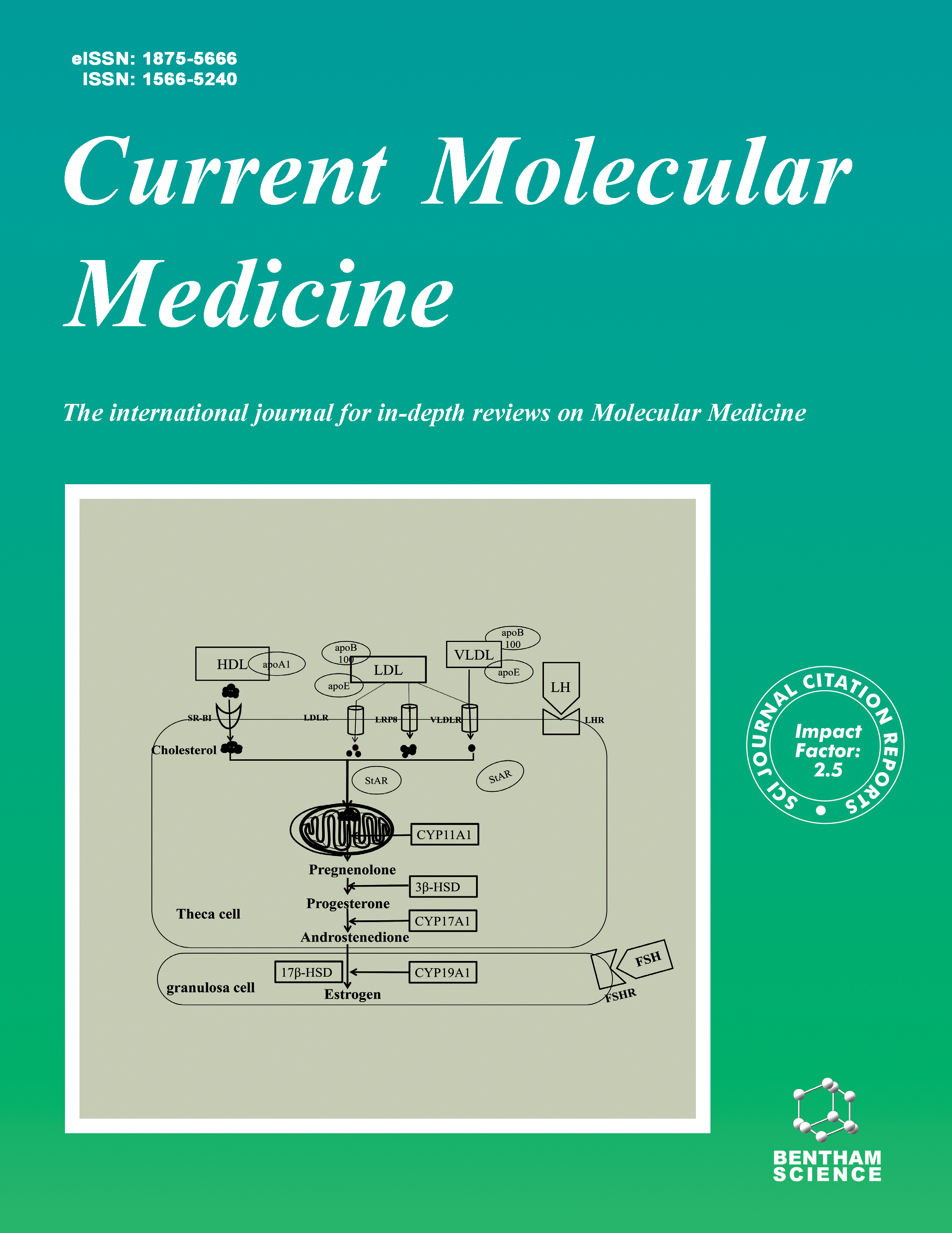
Current Molecular Medicine - Volume 9, Issue 7, 2009
Volume 9, Issue 7, 2009
-
-
N-glycosylation Engineering of Biopharmaceutical Expression Systems
More LessAuthors: P. P. Jacobs and N. CallewaertN-glycosylation, the enzymatic coupling of oligosaccharides to specific asparagine residues of nascent polypeptide chains, is one of the most widespread post-translational modifications. Following transfer of an N-glycan precursor in the ER, this structure is further modified by a number of glycosidases and glycosyltransferases in the ER and the Golgi complex. The processing reactions occurring in the ER are highly conserved between lower and higher eukaryotes. In contrast, the reactions that take place in the Golgi complex are species- and cell type-specific. Due to its non-template driven nature, glycoproteins typically occur as a mixture of glycoforms. Since N-glycans influence circulation half-life, tissue distribution, and biological activity each glycoform has its own pharmacokinetic, pharmacodynamic and efficacy profile. Moreover, modification of glycoproteins with non-human oligosaccharides can result in undesired immunogenicity. Therefore, engineering of the N-glycosylation pathway of most currently used heterologous protein expression systems (bacteria, mammalian cells, insect cells, yeasts and plants) is actively pursued by several academic and industrial laboratories. These research efforts are in the first place directed at humanizing the Nglycosylation pathway and eliminating immunogenic glycotopes. Moreover, one wants to establish new structure-function relationships of different glycoforms, which helps to decreasing the complexity of the Nglycan repertoire towards one defined N-glycan structure. In this review, we discuss the most important recent milestones in the glycoengineering field.
-
-
-
Mining the Genome for Susceptibility to Complex Neurological Disorders
More LessAuthors: A. K. Gubitz and K. GwinnGenome-wide association studies (GWAS) have become increasingly widely used to determine regions of the genome which may contain loci influencing the risk of neurological disorders. While linkage studies have identified genes that cause a number of Mendelian disorders, linkage analysis is less well suited for the more common complex disorders. This has led to the widespread use of GWAS for that purpose. Here we present and discuss several of the major extant GWAS in neurological disorders, their limitations, and implications of findings to date.
-
-
-
B-Type Natriuretic Peptide: Endogenous Regulator of Myocardial Structure, Biomarker and Therapeutic Target
More LessAuthors: Rebecca H. Ritchie, Anke C. Rosenkranz and David M. KayeB-type natriuretic peptide (BNP), initially identified in brain tissues, is now recognized as a key cardiac hormone. Numerous studies over the last decade have demonstrated that both exogenous and endogenous BNP prevent left ventricular (LV) hypertrophy in experimental settings, largely via activation of particulate guanylyl cyclase (pGC)-coupled receptors. BNP represents somewhat of a paradox, in that upregulation of BNP expression is widely used as a diagnostic marker for LV hypertrophy, diastolic dysfunction and heart failure in the clinic. We and others have postulated that BNP serves as an endogenous brake on the LV myocardium, seeking to curb the runaway train of signaling pathways that drive the progression from LV hypertrophy though remodeling, heart failure and death. This review summarizes the mechanisms of BNP's antihypertrophic actions, the role for cyclic GMP-mediated inhibition of pro-hypertrophic signaling, and BNP's impact on LV function. The improved understanding of the mechanisms of BNP regulation of LV hypertrophy and function that has emerged from both the experimental and clinical experience with this peptide provides new insight into the potential that BNP pharmacotherapy still offers for patients with LV hypertrophy.
-
-
-
Prion Protein Misfolding
More LessAuthors: L. Kupfer, W. Hinrichs and M. H. GroschupThe crucial event in the development of transmissible spongiform encephalopathies (TSEs) is the conformational change of a host-encoded membrane protein - the cellular PrPC - into a disease associated, fibril-forming isoform PrPSc. This conformational transition from the α8-helix-rich cellular form into the mainly β- sheet containing counterpart initiates an 'autocatalytic' reaction which leads to the accumulation of amyloid fibrils in the central nervous system (CNS) and to neurodegeneration, a hallmark of TSEs. The exact molecular mechanisms which lead to the conformational change are still unknown. It also remains to be brought to light how a polypeptide chain can adopt at least two stable conformations. This review focuses on structural aspects of the prion protein with regard to protein-protein interactions and the initiation of prion protein misfolding. It therefore highlights parts of the protein which might play a notable role in the conformational transition from PrPC to PrPSc and consequently in inducing a fatal chain reaction of protein misfolding. Furthermore, features of different proteins, which are able to adopt insoluble fibrillar states under certain circumstances, are compared to PrP in an attempt to understand the unique characteristics of prion diseases.
-
-
-
Role of β7 Integrins in Intestinal Lymphocyte Homing and Retention
More LessAuthors: G. Gorfu, J. Rivera-Nieves and K. LeyLymphocytes involved in intestinal immune response are found in organized immune inductive sites of the gut-associated lymphoid tissues (GALT) such as Peyer's patches (PP), mesenteric lymph nodes (MLN) and diffuse effector sites of gut epithelium and lamina propria (LP). β7 integrins are responsible for efficient trafficking and retention of lymphocytes in these sites. Naïve and effector lymphocytes use α4β7 integrin to extravasate from blood to gut mucosal tissues of GALT, MLN and LP via interactions with Mucosal Addressin Cell Adhesion Molecule-1 (MAdCAM-1). The αEβ7 integrin facilitates retention of effector and memory lymphocytes in the gut epithelial layer via interactions with E-cadherin. Mucosal dendritic cells (DCs) regulate the expression of the gut homing receptors α4β7 integrin and the chemokine receptor CCR9 on activated effector and regulatory lymphocytes in a retinoic acid-dependent manner. CD103 (αE integrin) identifies a subset of mucosal DCs in MLN and small intestine LP that have an enhanced ability to induce gut-tropic receptors on responding lymphocytes. The interactions between β7 integrin and their ligands are also implicated in the pathogenesis and progression of inflammatory bowel diseases (IBDs), intestinal parasitic infections and graft-versus-host diseases. During intestinal inflammation, β7 integrin-dependent and - independent pathways contribute to lymphocytes recruitment to the intestinal tissues and disease pathogenesis. Recent works have explored the potential of therapeutic targeting of α4 and β7 integrins in IBDs. Here, we review the current understanding of the role of β7 integrins in intestinal lymphocyte trafficking and retention in health and disease.
-
-
-
Spinal Muscular Atrophy: Molecular Mechanisms
More LessAuthors: M. A. Farrar, H. M. Johnston, P. Grattan-Smith, A. Turner and M. C. KiernanSpinal muscular atrophy (SMA) is a relatively common autosomal recessive neuromuscular disorder characterised by muscle weakness and atrophy due to degeneration of motor neurons of the spinal cord and cranial motor nuclei. The clinical phenotype incorporates a wide spectrum. No effective treatment is currently available and patients may experience severe physical disability which is often life limiting. The most common type of SMA is caused by homozygous disruption of the survival motor neuron 1 (SMN1) gene by deletion, conversion or mutation and results in insufficient levels of survival motor neuron (SMN) protein in motor neurons. While diagnosis is usually achieved by genetic testing, an illustrative clinical case is described that highlights the molecular and diagnostic complexities. While there is an emerging picture concerning the function of the SMN protein and the molecular pathophysiological mechanisms underpinning the disease, a number of substantial issues remain unresolved. The selective vulnerability of the motor neuron and the site and timing of the primary pathogenesis are not yet determined. Utilising the organisation of the SMN genomic region, recent advances have identified a number of potential therapeutic targets. As such, this review incorporates discussion of the clinical manifestations, molecular genetics, diagnosis, mechanisms of disease pathogenesis and development of novel treatment strategies.
-
-
-
HSP27: Mechanisms of Cellular Protection Against Neuronal Injury
More LessAuthors: R. A. Stetler, Y. Gao, A. P. Signore, G. Cao and J. ChenThe heat shock protein (HSP) family has long been associated with a generalized cellular stress response, particularly in terms of recognizing and chaperoning misfolded proteins. While HSPs in general appear to be protective, HSP27 has recently emerged as a particularly potent neuroprotectant in a number of diverse neurological disorders, ranging from ALS to stroke. Although its robust protective effect on a number of insults has been recognized, the mechanisms and regulation of HSP27's protective actions are still undergoing intense investigation. On the basis of recent studies, HSP27 appears to have a dynamic and diverse range of function in cellular survival. This review provides a forum to compare and contrast recent literature exploring the protective mechanism and regulation of HSP27, focusing on neurological disorders in particular, as they represent a range from protein aggregate-associated diseases to acute stress.
-
-
-
Hedgehog Target Genes: Mechanisms of Carcinogenesis Induced by Aberrant Hedgehog Signaling Activation
More LessHedgehog signaling is aberrantly activated in glioma, medulloblastoma, basal cell carcinoma, lung cancer, esophageal cancer, gastric cancer, pancreatic cancer, breast cancer, and other tumors. Hedgehog signals activate GLI family members via Smoothened. RTK signaling potentiates GLI activity through PI3KAKT- mediated GSK3 inactivation or RAS-STIL1-mediated SUFU inactivation, while GPCR signaling to Gs represses GLI activity through adenylate cyclase-mediated PKA activation. GLI activators bind to GACCACCCA motif to regulate transcription of GLI1, PTCH1, PTCH2, HHIP1, MYCN, CCND1, CCND2, BCL2, CFLAR, FOXF1, FOXL1, PRDM1 (BLIMP1), JAG2, GREM1, and Follistatin. Hedgehog signals are finetuned based on positive feedback loop via GLI1 and negative feedback loop via PTCH1, PTCH2, and HHIP1. Excessive positive feedback or collapsed negative feedback of Hedgehog signaling due to epigenetic or genetic alterations leads to carcinogenesis. Hedgehog signals induce cellular proliferation through upregulation of N-Myc, Cyclin D/E, and FOXM1. Hedgehog signals directly upregulate JAG2, indirectly upregulate mesenchymal BMP4 via FOXF1 or FOXL1, and also upregulate WNT2B and WNT5A. Hedgehog signals induce stem cell markers BMI1, LGR5, CD44 and CD133 based on cross-talk with WNT and/or other signals. Hedgehog signals upregulate BCL2 and CFLAR to promote cellular survival, SNAI1 (Snail), SNAI2 (Slug), ZEB1, ZEB2 (SIP1), TWIST2, and FOXC2 to promote epithelial-to-mesenchymal transition, and PTHLH (PTHrP) to promote osteolytic bone metastasis. KAAD-cyclopamine, Mu-SSKYQ-cyclopamine, IPI-269609, SANT1, SANT2, CUR61414 and HhAntag are small-molecule inhibitors targeted to Smoothened, GANT58, GANT61 to GLI1 and GLI2, and Robotnikinin to SHH. Hedgehog signaling inhibitors should be used in combination with RTK inhibitors, GPCR modulators, and/or irradiation for cancer therapy.
-
-
-
Effects of α-Crystallin on Lens Cell Function and Cataract Pathology
More LessThe development of cataracts is a debilitating eye condition which is common in elderly patients and afflicts millions worldwide. Cataracts result from the deposition of aggregated proteins in the eye which causes clouding of the lens, light scattering, and obstruction of vision. Non-syndromic, hereditary human cataract development is linked to point mutations in the CRYAA and CRYAB genes which encode αA and αB-crystallin. The α-crystallins are small heat shock proteins which play central roles in maintaining lens transparency and refractive properties. The discovery in 1992 that these proteins possess chaperone-like activity has led most researchers to focus on the ability of α-crystallins to prevent protein aggregation in vitro. While the ability of α- crystallins to efficiently trap aggregation-prone denatured proteins in vitro is thought to delay the development of age-related cataracts in vivo, α-crystallins have additional functions which may also contribute to cataract pathology. In addition to chaperone activity, α-crystallins are known to protect cells from stress-induced apoptosis, regulate cell growth, and enhance genomic stability. They also physically and functionally interact with both the cell membrane and cytoskeleton. Functional changes in α-crystallin have been shown to modify membrane and cell-cell interactions and lead to lens cell pathology in vivo. This article focuses on the multiple diverse roles of αA-crystallin in the maintenance of lens function and cataract development in vivo.
-
-
-
Dendritic Cells: A New Player in Osteoimmunology
More LessAuthors: M. Alnaeeli and Y. -T. A. TengRecent studies have suggested that the dys-regulated progressive immune responses in some inflammatory conditions can lead to significantly increased osteoclasts (OC) frequency and activity associated with active bone destruction; termed inflammation-induced bone loss. Among the inflammatory infiltrates, monocytes/macrophages (Mo/MQ), T and B cells, have been well studied and documented as central players in osteoimmunological interactions (osteoimmunology: is an interdisciplinary field linking the immune and skeletal systems). We and others investigated the role(s) of dendritic cells (DC) during inflammation-induced osteoclastogenesis and bone loss. In addition to their innate effector functions, DC are potent professional antigen-presenting cells (APC) involved in triggering and orchestrating adaptive immunity, thereby implicated as potential osteo-immune players. Herein, bone remodeling and DC's biology including their development and functions are reviewed along with the contribution of DC at the crossroad of the osteo-immune interface during the process of inflammation-induced osteoclastogenesis. Furthermore, we provide a summary of recent progress, and discuss a proposed alternative mechanism underlying inflammation-induced bone loss. Understanding the cellular and molecular mechanisms regulating DC's roles in inflammation-induced osteoclastogenesis and bone loss might benefit future treatment approaches, especially if targeting DC can be translated into therapeutic strategies to ameliorate both tissue inflammation and bone destruction during disease progression associated with inflammatory bone diseases.
-
-
-
Cosignaling Molecules Around LIGHT-HVEM-BTLA: From Immune Activation to Therapeutic Targeting
More LessAuthors: Christine Pasero, Alemseged Truneh and Daniel OliveThe regulation of the immune system at the cell surface is primarily controlled by two families of cosignaling molecules: the immunoglobulin (Ig) superfamily, or ≪CD28 and B7 family≫, and the tumor necrosis factor receptor (TNFR) family. Here, we summarized the principal structural and functional characteristics of both families. In this respect, the interaction between HVEM, a TNF receptor, and BTLA, an Ig family member, has provided a new perspective and an additional level of complexity in the crosstalk between these two regulatory systems. This review will present a summary of the recent advances in the immunobiology of the LIGHT-HVEM-LTβR-BTLA network. The LIGHT-HVEM-BTLA system has emerged as a major regulator of immune responses and lymphocyte activation, whereas LIGHT-LTβR participates in lymphoid tissue development and cell death. Moreover, recent studies have provided encouraging new insights into the roles of the LIGHT-HVEM-LTβR-BTLA axis as a potential target for controlling anti-tumor responses.
-
Volumes & issues
-
Volume 25 (2025)
-
Volume 24 (2024)
-
Volume 23 (2023)
-
Volume 22 (2022)
-
Volume 21 (2021)
-
Volume 20 (2020)
-
Volume 19 (2019)
-
Volume 18 (2018)
-
Volume 17 (2017)
-
Volume 16 (2016)
-
Volume 15 (2015)
-
Volume 14 (2014)
-
Volume 13 (2013)
-
Volume 12 (2012)
-
Volume 11 (2011)
-
Volume 10 (2010)
-
Volume 9 (2009)
-
Volume 8 (2008)
-
Volume 7 (2007)
-
Volume 6 (2006)
-
Volume 5 (2005)
-
Volume 4 (2004)
-
Volume 3 (2003)
-
Volume 2 (2002)
-
Volume 1 (2001)
Most Read This Month


