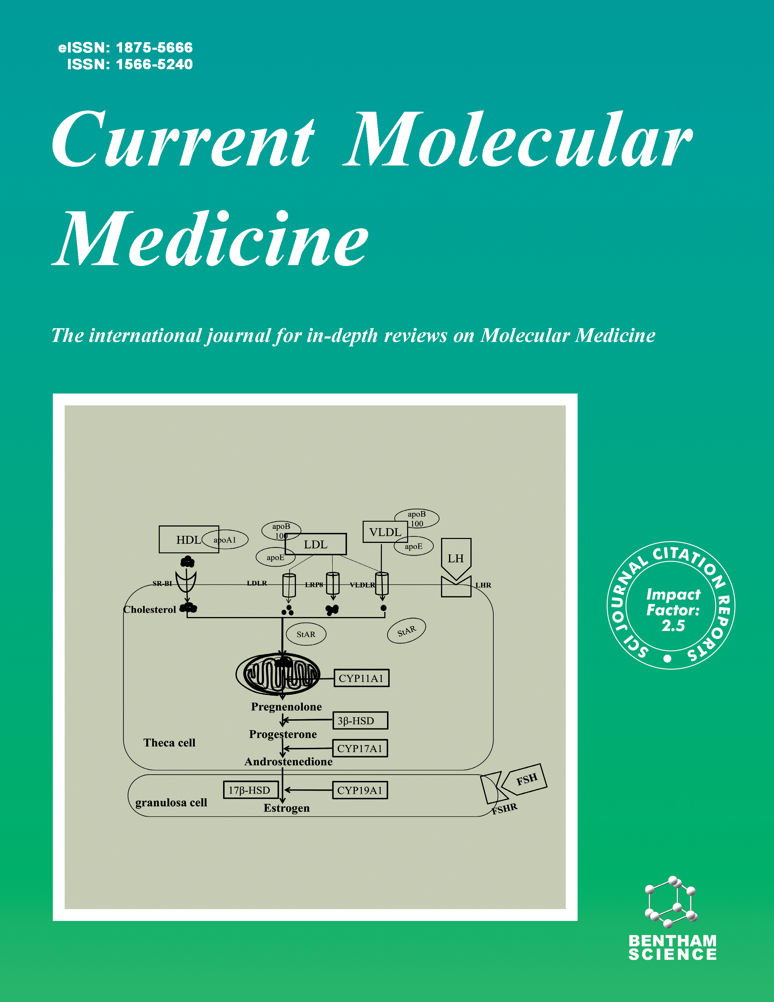
Current Molecular Medicine - Volume 8, Issue 8, 2008
Volume 8, Issue 8, 2008
-
-
MicroRNAs in Organogenesis and Disease
More LessAuthors: Naisana S. Asli, Mara E. Pitulescu and Michael KesselLarge numbers and quantities of different, small RNA molecules are present in the cytoplasm of animal and plant cells. One subclass of these molecules is represented by the noncoding microRNAs. Since their discovery in the 1990s a multitude of basic information has accumulated, which has identified their function in post-transcriptional control, either via degradation or translational inhibition of target mRNAs. This function is in most of the cases a finetuning of gene expression, working in parallel with transcriptional regulatory processes. MicroRNA expression profiles are highly dynamic during embryonic development and in adulthood. Misexpression of microRNAs can perturb embryogenesis, organogenesis, tissue homeostasis and the cell cycle. Evidence from gain- and loss-of function studies indicates roles for microRNAs in pathophysiologic states including cardiac hypertrophy, muscle dystrophy, hepatitis infection, diabetes, Parkinson syndrome, hematological malignancies and other types of cancer. In this review, we focus on studies addressing the role of various microRNAs in heart, muscle, liver, pancreas, central nervous system, and hematopoiesis.
-
-
-
Molecular Imaging of Brain Tumors Personal Experience and Review of the Literature
More LessAuthors: Bernhard J. Schaller, Jan F. Cornelius, Nora Sandu and Michael BuchfelderNon-invasive energy metabolism measurements in brain tumors in vivo are now performed widely as molecular imaging by positron emission tomography. This capability has developed from a large number of basic and clinical science investigations that have cross fertilized one another. Apart from precise anatomical localization and quantification, the most intriguing advantage of such imaging is the opportunity to investigate the time course (dynamics) of disease-specific molecular events in the intact organism. Most importantly, molecular imaging represents a key-technology in translational research, helping to develop experimental protocols that may later be applied to human patients. Common clinical indications for molecular imaging of primary brain tumors therefore contain (i) primary brain tumor diagnosis, (ii) identification of the metabolically most active brain tumor reactions (differentiation of viable tumor tissue from necrosis), and (iii) prediction of treatment response by measurement of tumor perfusion, or ischemia. The key-question remains whether the magnitude of biochemical alterations demonstrated by molecular imaging reveals prognostic value with respect to survival. Molecular imaging may identify early disease and differentiate benign from malignant lesions. Moreover, an early identification of treatment effectiveness could influence patient management by providing objective criteria for evaluation of therapeutic strategies for primary brain tumors. Specially, its novel potential to visualize metabolism and signal transduction to gene expression is used in reporter gene assays to trace the location and temporal level of expression of therapeutic and endogenous genes. The authors present here illustrative data of PET imaging: the thymidine kinase gene expression in experimentally transplanted F98 gliomas in cat brain indicates, that [18F]FHBG visualizes cells expressing TK-GFP gene in transduced gliomas as well as quantities and localizes transduced HSV-1-TK expression if the blood brain barrier is disrupted. The higher uptake of [18F]FLT in the wild-type compared to the transduced type may demonstrate the different doubling time of both tumor tissues suggesting different cytosolic thymidine kinase activity. Molecular imaging probes are developed to image the function of targets without disturbing them or as drug in oder to modify the target's function. This is transfer of gene therapy's experimental knowledge into clinical applications. Molecular imaging closes the gap between in vitro to in vivo integrative biology of disease.
-
-
-
IGF Signaling Pathway as a Selective Target of Familial Breast Cancer Therapy
More LessAuthors: Vivek Shukla, Xavier Coumoul, Athanassios Vassilopoulos and Chu-Xia DengHereditary breast cancers affect women who have an increased risk of developing tumors because of a familial history. In most cases, they can be attributed to mutations in the breast cancer associated gene 1 and 2 (BRCA1 and BRCA2). Recent studies have demonstrated a link between the insulin-like growth factor (IGF) signaling pathway and familial breast cancer incidence. IGF and IGF receptors represent a family of biological growth factors and transducers, which have been involved in both physiological and pathological processes. It has been shown that BRCA1 regulates expression of several members of the IGF family. Here, we will examine our understanding of the functions of IGF/IGF-receptor signaling, the development of new inhibitors of this pathway and the related mechanisms of familial breast cancer formation.
-
-
-
Caveolae and Caveolins in the Respiratory System
More LessAuthors: Reinoud Gosens, Mark Mutawe, Sarah Martin, Sujata Basu, Sophie T. Bos, Thai Tran and Andrew J. HalaykoCaveolae are flask-shaped invaginations of the plasma membrane that are present in most structural cells. They owe their characteristic Ω-shape to complexes of unique proteins, the caveolins, which indirectly tether cholesterol and sphingolipid-enriched membrane microdomains to the cytoskeleton. Caveolins possess a unique scaffolding domain that anchors receptors, ion channels, second messenger producing enzymes, and effector kinases, thereby sequestering them to caveolae, and modulating cellular signaling and vesicular transport. The lungs express numerous caveolae and high levels of caveolins; therefore they likely play an important role in lung physiology. Indeed, recent and ongoing studies indicate important roles for caveolae and caveolins in the airway epithelium, airway smooth muscle, airway fibroblasts, airway inflammatory cells and the pulmonary vasculature. We review the role of caveolae and caveolins in lung cells and discuss their involvement in cellular signaling associated with asthma, COPD, lung cancer, idiopathic pulmonary fibrosis and pulmonary vascular defects.
-
-
-
Decreased Vascular Repair and Neovascularization with Ageing: Mechanisms and Clinical Relevance with an Emphasis on Hypoxia- Inducible Factor-1
More LessAuthors: Michel R. Hoenig, Cesario Bianchi, Anthony Rosenzweig and Frank W. SellkeAgeing is associated with endothelial dysfunction, decreased endothelial progenitor cell (EPC) function and mobilization. These defects culminate in a decreased capacity for neovascularization in the aged. Multiple lines of evidence suggest that defective neovascularization with ageing is related to depressed signaling by hypoxia inducible factor-1 (HIF-1). HIF-1, the master regulator or neovascularization, regulates the expression of vascular endothelial growth factor (VEGF), stromal cell-derived factor-1 (SDF-1) and CXC chemokine Receptor-4 (CXCR4). Given that the SDF-1/CXCR4 axis is a crucial regulator of progenitor cell function and homing, the ramifications of depressed HIF-1 signaling with age include depressed vascular repair, neovascularization and wound healing. We review the literature showing the depression of these processes with age and discuss the relevance of these findings to several clinical contexts. Further, the effects of age on EPC number, function and mobilization are related to the age-related decline in HIF-1 signaling. We suggest that exercise, Cobalt compounds or hydralazine may reverse the age-related decline by up-regulating HIF-1- mediated signaling.
-
-
-
Do Human Lipoxygenases have a PDZ Regulatory Domain?
More LessHuman lipoxygenases and products of their catalytic reaction have a well established connection to many human diseases. Despite their importance in inflammation, cancer, cardiorenal and other ailments the drug development is impaired by the lack of structural details to understand their intricate specificity and function in molecular and cellular signaling. The major effort so far has been directed towards understanding the determinants of their specificity and inhibition of their active site with the iron cofactor. Their structure is believed to consist of only two domains: one regulatory - a β-sandwich, important for membrane binding, and one, mostly helical, catalytic domain. Although recently published cohort studies on single nucleotide polymorphism and occurrence of diseases, SAXS analysis and new biochemical data throw new light on lipoxygenase suggesting symbiosis of regulatory functions with an allosteric mechanism and more flexible structure than anticipated. The goal of this brief review is to direct an attention to the structural features of an anticipated topology and stimulate discussion/research to prove or disapprove our hypothesis that lipoxygenases may possess about ∼110 amino acids PDZ-like fragments of functional importance. If they do have a second regulatory domain, it might help to explain their association with other molecules, role in signaling pathways and present a new avenue to explore the regulation of their behavior, and thus intervention in the course of diseases.
-
-
-
Integrated Genomic and Pharmacological Approaches to Identify Synthetic Lethal Genes as Cancer Therapeutic Targets
More LessAuthors: Shinji Mizuarai, Hiroki Irie, Dennis M. Schmatz and Hidehito KotaniVarious types of cancers are generated through mutations or dysregulations of oncogenes/tumor suppressor genes involved in cell cycles and signaling transduction pathways. To identify cancer therapeutic targets whose inhibition selectively kills cancer cells, synthetic lethal screening is being developed to identify genes whose intervention suppresses tumor progression only when combined with the dysregulation of the genes. The recent emergence of genomic technologies, including microarray, RNA interference and chemogenomics, provides platforms to realize this concept. This review introduces the research that could successfully identify synthetic lethal genes in cancer cells harboring major gene alterations such as p53, RB, K-Ras, or Myc. We also illustrate remarkable candidate targets that were identified by synthetic lethal screening to find chemosensitizers for paclitaxel and cisplatin. Next, we introduce the chemogenomics approaches that explore chemical compounds that exhibit synthetic lethality to cancer gene alterations. Although the synthetic lethal compounds are of great interest in terms of cancer drug development, a method of identifying target proteins for the phenotypic compounds has been elusive. Finally, we demonstrate several noteworthy techniques to identify target proteins for the compounds: a Connectivity Map that compares expression profiles of compoundtreated cells by pattern-matching algorithms; an siRNA/compound co-treatment assay to find enhancer genes for the phenotypes of compounds; and a state-of-the-art proteomics approach that modifies classical compound- immobilized affinity chromatography. The integration of genomic and pharmacological analyses would significantly accelerate the identification of cancer-specific synthetic lethal targets.
-
-
-
CD44 and EpCAM: Cancer-Initiating Cell Markers
More LessEmbryonic stem cells are immortal, can self renew, and differentiate into all cells of the body. The adult organism maintains adult stem cells in regenerative organs that can differentiate into all cells of the respective organ. Virchow's hypothesis that cancer may arise from embryonic-like cells has received strong support, as it was demonstrated that tumors contain few cells, known as cancer stem or cancer-initiating cells (CIC), that account for primary and metastatic tumor growth. CIC are mostly defined by expression of CIC-markers that are associated and correlated with the potential of CIC to grow in xenogeneic mice. CIC marker profiles have been elaborated for many tumors, with several markers as CD24, CD44, CD133, CD166, EpCAM, and some integrins, being expressed by tumors of different histological type. Their function in promoting CIC maintenance and activity is largely unknown. The fate of stem cells, determined by their position, is minutely regulated by few adjacent cells creating a niche. CIC also require a niche, mostly for settlement and growth in distant organs. This so called pre-metastatic niche is initiated by the primary tumor before metastasizing cell arrival. How do CIC prepare the pre-metastatic niche? Cancer cells secrete a matrix that serves a cross-talk with surrounding tissues. Additionally, cancer cells can abundantly deliver exosomes, which function as long-distance intercellular communicators. Studies on a rat pancreatic adenocarcinoma support our hypothesis that tumorderived matrix and exosomes are the main actors in forming the pre-metastatic niche with CIC markers being engaged in matrix preparation and/or exosome delivery.
-
-
-
Menin, Histone H3 Methyltransferases, and Regulation of Cell Proliferation: Current Knowledge and Perspective
More LessAuthors: Xinjiang Wu and Xianxin HuaMenin is a tumor suppressor encoded by the MEN1 gene that is mutated in patients with an inherited syndrome, multiple endocrine neoplasia type 1 (MEN1). Loss of menin has potent impact on proliferation of endocrine and non-endocrine cells. However, until recently little has been known as to how menin regulates cell proliferation. Rapid research progress in the past several years suggests that menin represses proliferation of endocrine cells yet promotes proliferation in certain types of leukemia cells via interacting with various transcriptional regulators. Menin interacts with histone H3 methyltransferases such as MLL (mixed lineage leukemia) protein. Increasing evidence has linked the biological function of menin to epigenetic histone modifications, control of the pattern of gene expression, and regulation of cell proliferation in a cell type-specific manner. In light of these recent findings, an emerging model suggests that menin is a crucial regulator of histone modifiers by acting as a scaffold protein to coordinate gene transcription and cell proliferation in a cell contextdependent manner. This recent progress unravels the coordinating role of menin in epigenetics and regulation of cell cycle, providing novel insights into understanding regulation of beta cell functions and diabetes, as well as the development and therapy of endocrine tumors and leukemia.
-
-
-
Hypogonadotrophic Hypogonadism in Type 2 Diabetes, Obesity and the Metabolic Syndrome
More LessAuthors: Paresh Dandona, Sandeep Dhindsa, Ajay Chaudhuri, Vishal Bhatia, Shehzad Topiwala and Priya MohantyRecent work shows a high prevalence of low testosterone and inappropriately low LH and FSH concentrations in type 2 diabetes. This syndrome of hypogonadotrophic hypogonadism (HH) is associated with obesity, and other features of the metabolic syndrome (obesity and overweight, hypertension and hyperlipidemia) in patients with type 2 diabetes. However, the duration of diabetes or HbA1c were not related to HH. Furthermore, recent data show that HH is also observed frequently in patients with the metabolic syndrome without diabetes but is not associated with type 1 diabetes. Thus, HH appears be related to the two major conditions associated with insulin resistance: type 2 diabetes and the metabolic syndrome. CRP concentrations have been shown to be elevated in patients with HH and are inversely related to plasma testosterone concentrations. This inverse relationship between plasma free testosterone and CRP concentrations in patients with type 2 diabetes suggests that inflammation may play an important role in the pathogenesis of this syndrome. This is of interest since inflammatory mechanisms may have a cardinal role in the pathogenesis of insulin resistance. It is relevant that in the mouse, deletion of the insulin receptor in neurons leads to HH in addition to a state of systemic insulin resistance. It has also been shown that insulin facilitates the secretion of gonadotrophin releasing hormone (GnRH) from neuronal cell cultures. Thus, HH may be the result of insulin resistance at the level of the GnRH secreting neuron. Low testosterone concentrations in type 2 diabetic men have also been related to a significantly lower hematocrit and thus to an increased frequency of mild anemia. Low testosterone concentrations are also related to an increase in total and regional adiposity, and to lower bone density. This review discusses these issues and attempts to make the syndrome relevant as a clinical entity. Clinical trials are required to determine whether testosterone replacement alleviates symptoms related to sexual dysfunction, and features of the metabolic syndrome, insulin resistance and inflammation.
-
-
-
The Expanding Role of APRIL in Cancer and Immunity
More LessAuthors: Lourdes Planelles, Jan P. Medema, Michael Hahne and Gijs HardenbergProteins of the tumour necrosis factor (TNF) family are implicated in the regulation of essential cell processes such as proliferation, differentiation, survival and cell death. Altered expression of TNF family members is often associated with pathological conditions such as autoimmune disease and cancer. The TNF-like ligand APRIL (A PRoliferation Inducing Ligand), first described in 1998, was named for its capacity to stimulate tumour cell proliferation in vitro. APRIL expression was initially reported in haematopoietic cells in physiological conditions, and it is overexpressed in certain tumour tissues. APRIL is now known to be involved in activation and immune responses of B cells, as well as in B cell malignancies. This review focuses on recent advances in understanding APRIL and its receptors in physiology and tumour pathology, including the accumulating evidence that specific Toll-like receptor ligands can trigger APRIL-mediated responses, and the identification of new sources of APRIL such as epithelial cells and tumour- infiltrating neutrophils.
-
-
-
Posttranscriptional Regulation of p53 and its Targets by RNABinding Proteins
More LessAuthors: Jin Zhang and Xinbin Chenp53 tumor suppressor plays a pivotal role in maintaining genomic integrity and preventing cancer development. The importance of p53 in tumor suppression is illustrated by the observation that about 50% human tumor cells have a dysfunctional p53 pathway. Although it has been well accepted that the activity of p53 is mainly controlled through post-translational modifications, recent studies have revealed that posttranscriptional regulations of p53 by various RNA-binding proteins also play a crucial role in modulating p53 activity and its downstream targets.
-
Volumes & issues
-
Volume 25 (2025)
-
Volume 24 (2024)
-
Volume 23 (2023)
-
Volume 22 (2022)
-
Volume 21 (2021)
-
Volume 20 (2020)
-
Volume 19 (2019)
-
Volume 18 (2018)
-
Volume 17 (2017)
-
Volume 16 (2016)
-
Volume 15 (2015)
-
Volume 14 (2014)
-
Volume 13 (2013)
-
Volume 12 (2012)
-
Volume 11 (2011)
-
Volume 10 (2010)
-
Volume 9 (2009)
-
Volume 8 (2008)
-
Volume 7 (2007)
-
Volume 6 (2006)
-
Volume 5 (2005)
-
Volume 4 (2004)
-
Volume 3 (2003)
-
Volume 2 (2002)
-
Volume 1 (2001)
Most Read This Month


