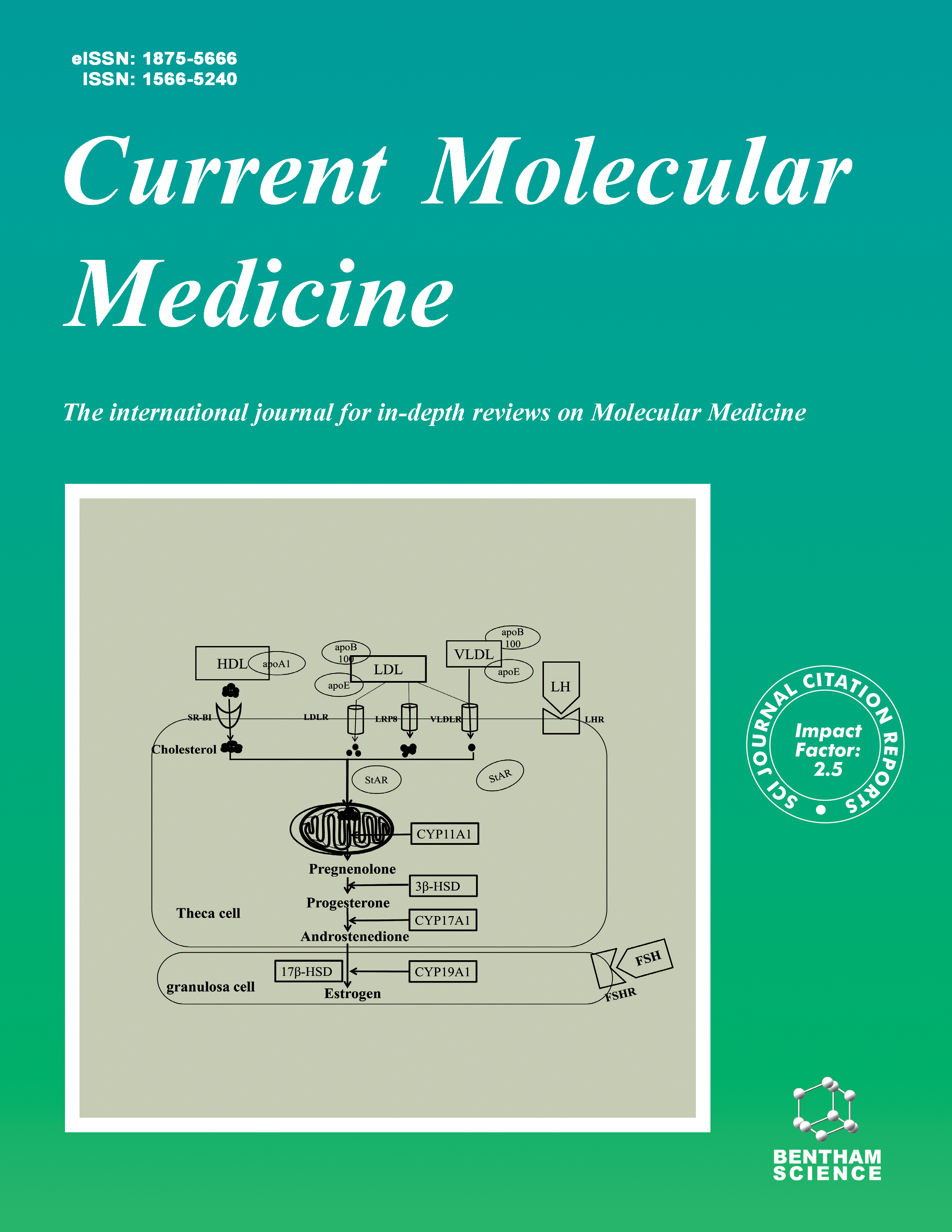
Current Molecular Medicine - Volume 4, Issue 2, 2004
Volume 4, Issue 2, 2004
-
-
Excitotoxic Versus Apoptotic Mechanisms of Neuronal Cell Death in Perinatal Hypoxia / Ischemia
More LessAuthors: Chainllie Young, Tatyana Tenkova, Krikor Dikranian and John W. OlneyHypoxic / / ischemic (H / I)neuronal degeneration in the developing central nervous system (CNS)is mediated by an excitotoxic mechanism,and it has also been reported that an apoptosis mechanism is involved.However,there is much disagreement regarding how excitotoxic and apoptotic cell death processes relate to one another.Some authors believe that an excitotoxic stimulus directly triggers apoptotic cell death,but this interpretation is largely speculative at the present time.Our findings support the interpretation that excitotoxic and apoptotic neurodegeneration are two separate and distinct cell death processes that can be distinguished from one another by ultrastructural evaluation.Here we review evidence supporting this interpretation,including evidence that H / I in the developing CNS triggers two separate waves of neurodegeneration,the first being excitotoxic and the second being apoptotic.The first (excitotoxic)wave destroys neurons that would normally provide synaptic inputs or synaptic targets for the neurons that die in the second (apoptotic)wave.Since neurons,during the developmental period of synaptogenesis,are programmed to commit suicide if they fail to achieve normal connectivity,this explains why neuroapoptosis occurs following H / I in the developing CNS.However,it does not support the interpretation that H / I directly triggers apoptotic neurodegeneration.Rather,it documents that H / I directly triggers excitotoxic neurodegeneration,and apoptotic neurodegeneration ensues subsequently as the natural response of developing neurons to a specific kind of deprivation -loss of the ability to form normal synaptic connections.
-
-
-
Rethinking the Excitotoxic Ionic Milieu:The Emerging Role of Zn 2+ in Ischemic Neuronal Injury
More LessAuthors: S. L. Sensi and J.- M. JengZn 2+plays an important role in diverse physiological processes,but when released in excess amounts it is potently neurotoxic.In vivo trans-synaptic movement and subsequent post-synaptic accumulation of intracellular Zn 2+contributes to the neuronal injury observed in some forms of cerebral ischemia.Zn 2+may enter neurons through NMDA channels,voltage-sensitive calcium channels,Ca 2+- permeable AMPA / kainate (Ca-A / K)channels,or Zn 2+-sensitive membrane transporters.Furthermore, Zn 2+is also released from intracellular sites such as metallothioneins and mitochondria.The mechanisms by which Zn 2+exerts its potent neurotoxic effects involve many signaling pathways, including mitochondrial and extra-mitochondrial generation of reactive oxygen species (ROS)and disruption of metabolic enzyme activity,ultimately leading to activation of apoptotic and / or necrotic processes. As is the case with Ca 2+,neuronal mitochondria take up Zn 2+as a way of modulating cellular Zn 2+ homeostasis.However,excessive mitochondrial Zn 2+sequestration leads to a marked dysfunction of these organelles,characterized by prolonged ROS generation.Intriguingly,in direct comparison to Ca 2+,Zn 2+appears to induce these changes with a considerably greater degree of potency.These effects are particularly evident upon large (i.e.,micromolar)rises in intracellular Zn 2+concentration ([Zn 2+]i ),and likely hasten necrotic neuronal death.In contrast,sub-micromolar [Zn 2+]i increases promote release of pro-apoptotic factors,suggesting that different intensities of [Zn 2+]i load may activate distinct pathways of injury.Finally,Zn 2+homeostasis seems particularly sensitive to the environmental changes observed in ischemia,such as acidosis and oxidative stress,indicating that alterations in [Zn 2+]i may play a very significant role in the development of ischemic neuronal damage.
-
-
-
White Matter Injury Mechanisms
More LessWhite matter of the brain and spinal cord is susceptible to anoxia,ischemia,trauma and autoimmune attack.Irreversible injury to this tissue can have serious consequences for the overall function of the CNS through disruption of signal transmission.Like neurons,central myelinated axons are critically dependent on a continuous supply of oxygen and glucose.Injury causes failure of the Na-K-ATPase and accumulation of axoplasmic Na through non-inactivating Na channels,which, together with membrane depolarization,promotes reverse Na-Ca exchange and axonal Ca overload.An equally important source of deleterious Ca originates from intracellular stores,released in part by a mechanism similar to “excitation-contraction coupling ” in muscle,,involving activation of ryanodine receptors by L-type Ca channels.Excitotoxic mechanisms also play an important role:glutamate released by reversal of Na-dependent glutamate transporters activates AMPA / kainate receptors to cause injury to glia and myelin.Excessive accumulation of cytosolic Ca in turn activates various Ca- dependent enzymes such as calpains,phospholipases and others resulting in irreversible injury. Reoxygenation paradoxically accelerates injury in many axons,and promotes cytoskeletal degradation. Blockers of voltage-gated Na channels represent an attractive therapeutic target because of their ability to simultaneously interfere indirectly with several Ca sourcing pathways.Alternatively,or additionally,AMPA / kainate receptor inhibition has also been shown to be neuroprotective in several white matter injury paradigms.In the clinical setting,optimal protection of the CNS as a whole in common disorders such as stroke,traumatic brain and spinal cord injury,will likely require combination therapy aimed at unique steps in gray and white matter regions,or intervention at common points in the injury cascades.
-
-
-
The Rise and Fall of NMDA Antagonists for Ischemic Stroke
More LessAuthors: L. Hoyte, P. A. Barber, A. M. Buchan and M. D. HillIt has long been accepted that high concentrations of glutamate can destroy neurons,and this is the basis of the theory of excitotoxicity during brain injury such as stroke.Glutamate N-methyl- D-aspartate (NMDA)receptor antagonists such as Selfotel,Aptiganel,Gavestinel and others failed to show neuroprotective efficacy in human clinical trials or produced intolerable central nervous system adverse effects.The failure of these agents has been attributed to poor studies in animal models and to poorly designed clinical trials.We also speculate that NMDA receptor anatagonism may have hindered endogenous mechanisms for neuronal survival and neuroregeneration.It remains to be proven in human stroke whether NMDA receptor antagonism can be neuroprotective.
-
-
-
Molecular Mechanisms Underlying Specificity of Excitotoxic Signaling in Neurons
More LessAuthors: Michelle M. Aarts and Michael TymianskiThe central role of glutamate receptors in mediating excitotoxic neuronal death in stroke, epilepsy and trauma has been well established.Glutamate is the major excitatory amino acid transmitter within the CNS and it 's signaling is mediated by a number of postsynaptic ionotropic and metabotropic receptors.Although calcium ions are considered key regulators of excitotoxicity,new evidence suggests that specific second messenger pathways rather than total Ca 2+load,are responsible for mediating neuronal degeneration.Glutamate receptors are found localized at the synapse within electron dense structures known as the postsynaptic density (PSD).Localization at the PSD is mediated by binding of glutamate receptors to submembrane proteins such as actin and PDZ containing proteins.PDZ domains are conserved motifs that mediate protein-protein interactions and self-association.In addition to glutamate receptors PDZ-containing proteins bind a multitude of intracellular signal molecules including nitric oxide synthase.In this way PDZ proteins provide a mechanism for clustering glutamate receptors at the synapse together with their corresponding signal transduction proteins.PSD organization may thus facilitate the individual neurotoxic signal mechanisms downstream of receptors during glutamate overactivity.Evidence exists showing that inhibiting signals downstream of glutamate receptors,such as nitric oxide and PARP-1 can reduce excitotoxic insult.Furthermore we have shown that uncoupling the interaction between specific glutamate receptors from their PDZ proteins protects neurons against glutamate-mediated excitotoxicity.These findings have significant implications for the treatment of neurodegenerative diseases using therapeutics that specifically target intracellular protein-protein interactions.
-
-
-
Mitochondrial Dysfunction and Glutamate Excitotoxicity Studied in Primary Neuronal Cultures
More LessPrimary dissociated neuronal cultures have been intensively exploited for the past 15 years as model systems to investigate excitotoxic neuronal degeneration.Even this simplified system contains a complex web of interactions between calcium homeostasis,ATP production and the generation and detoxification of reactive oxygen species.There is increasing realization that the mitochondrion occupies the center stage in these processes.This review covers the normal bioenergetics of the cultured neuron,the ways in which mitochondrial dysfunction impacts upon the ability of the neuron to withstand excitotoxic stress,the nature of the stresses imposed by NMDA receptor activation and possible molecular mechanisms of excitotoxic cell death.
-
-
-
Nitric Oxide and its Role in Ischaemic Brain Injury
More LessAuthors: Robert G. Keynes and John GarthwaiteThe role of the neural messenger nitric oxide (NO)in cerebral ischaemia has been investigated extensively in the past decade.NO may play either a protective or destructive role in ischaemia and the literature is plagued with contradictory findings.Working with NO presents many unique difficulties and here we review the potential artifacts that may have contributed to discrepancies and cause future problems for the unwary investigator.Recent evidence challenges the idea that NO from neurones builds up to levels (micromolar)sufficient to directly elicit cell death during the post-ischaemic period.Concomitantly,the case is strengthened for a role of NO in delayed death mediated post-ischaemia by the inducible NO synthase.Mechanistically it seems unlikely that NO is released in high enough quantities to inhibit respiration in vivo ;the formation of reactive nitrogen species,such as peroxynitrite,represents the more likely pathway to cell death.The protective and restorative properties of NO have become of increasing interest.NO from endothelial cells may,via stimulating cGMP production,protect the ischaemic brain by acutely augmenting blood flow,and by helping to form new blood vessels in the longer term (angiogenesis).Elevated cGMP production may also stop cells dying by inhibiting apoptosis and help repair damage by stimulating neurogenesis.In addition NO may act as a direct antioxidant and participate in the triggering of protective gene expression programmes that underlie cerebral ischaemic preconditioning.Better understanding of the molecular mechanisms by which NO is protective may ultimately identify new potential therapeutic targets.
-
-
-
Astrocyte Influences on Ischemic Neuronal Death
More LessAuthors: Raymond A. Swanson, Weihai Ying and Tiina M. KauppinenGlutamate excitotoxicity,oxidative stress,and acidosis are primary mediators of neuronal death during ischemia and reperfusion.Astrocytes influence these processes in several ways. Glutamate uptake by astrocytes normally prevents excitotoxic glutamate elevations in brain extracellular space,and this process appears to be a critical determinant of neuronal survival in the ischemic penumbra.Conversely,glutamate efflux from astrocytes by reversal of glutamate uptake, volume sensitive organic ion channels,and other routes may contribute to extracellular glutamate elevations.Glutamate activation of neuronal N-methyl-D-aspartate (NMDA)receptors is modulated by glycine and D-serine:both of these neuromodulators are transported by astrocytes,and D-serine production is localized exclusively to astrocytes.Astrocytes influence neuronal antioxidant status through release of ascorbate and uptake of its oxidized form,dehydroascorbate,and by indirectly supporting neuronal glutathione metabolism.In addition,glutathione in astrocytes can serve as a sink for nitric oxide and thereby reduce neuronal oxidant stress during ischemia.Astrocytes probably also influence neuronal survival in the post-ischemic period.Reactive astrocytes secrete nitric oxide, TNF α ,matrix metalloproteinases,and other factors that can contribute to delayed neuronal death,and facilitate brain edema via aquaporin-4 channels localized to the astrocyte endfoot-endothelial interface.On the other hand erythropoietin,a paracrine messenger in brain,is produced by astrocytes and upregulated after ischemia.Erythropoietin stimulates the Janus kinase-2 (JAK-2)and nuclear factor-kappaB (NF-kB)signaling pathways in neurons to prevent programmed cell death after ischemic or excitotoxic stress.Astrocytes also secrete several angiogenic and neurotrophic factors that are important for vascular and neuronal regeneration after stroke.
-
-
-
What have Genetically Engineered Mice Taught Us About Ischemic Injury?
More LessAuthors: Dong Liang, Ted M. Dawson and Valina L. DawsonStroke,is the third leading cause of death and disability in the Western world.Stroke refers to set of ischemic conditions resulting from the occlusion or hemorrhage of blood vessels supplying the brain.Loss of blood flow to the brain results in neuronal injury due to both oxygen and nutrient deprivation and the activation of injurious signal cascades.Ultimately cerebral ischemia results in death and dysfunction of brain cells,and neurological deficits that reflect the location and size of the compromised brain area.Injury due to ischemic stroke occurs by a highly choreographed series of complex spatial and temporal events that evolve over hours to days.These events involve complex interactions between fundamental cell injury mechanisms including excitotoxicity and ionic imbalance, oxidative and nitrosative stress,apoptotic-like cell death and inflammatory responses.Genetically engineered mice have been valuable tools to probe putative mechanisms of neuronal death and uncover potential strategies that might render neurons resistant to ischemic injury.Findings from experimental stroke studies in genetically engineered animals are discussed.
-
Volumes & issues
-
Volume 25 (2025)
-
Volume 24 (2024)
-
Volume 23 (2023)
-
Volume 22 (2022)
-
Volume 21 (2021)
-
Volume 20 (2020)
-
Volume 19 (2019)
-
Volume 18 (2018)
-
Volume 17 (2017)
-
Volume 16 (2016)
-
Volume 15 (2015)
-
Volume 14 (2014)
-
Volume 13 (2013)
-
Volume 12 (2012)
-
Volume 11 (2011)
-
Volume 10 (2010)
-
Volume 9 (2009)
-
Volume 8 (2008)
-
Volume 7 (2007)
-
Volume 6 (2006)
-
Volume 5 (2005)
-
Volume 4 (2004)
-
Volume 3 (2003)
-
Volume 2 (2002)
-
Volume 1 (2001)
Most Read This Month


