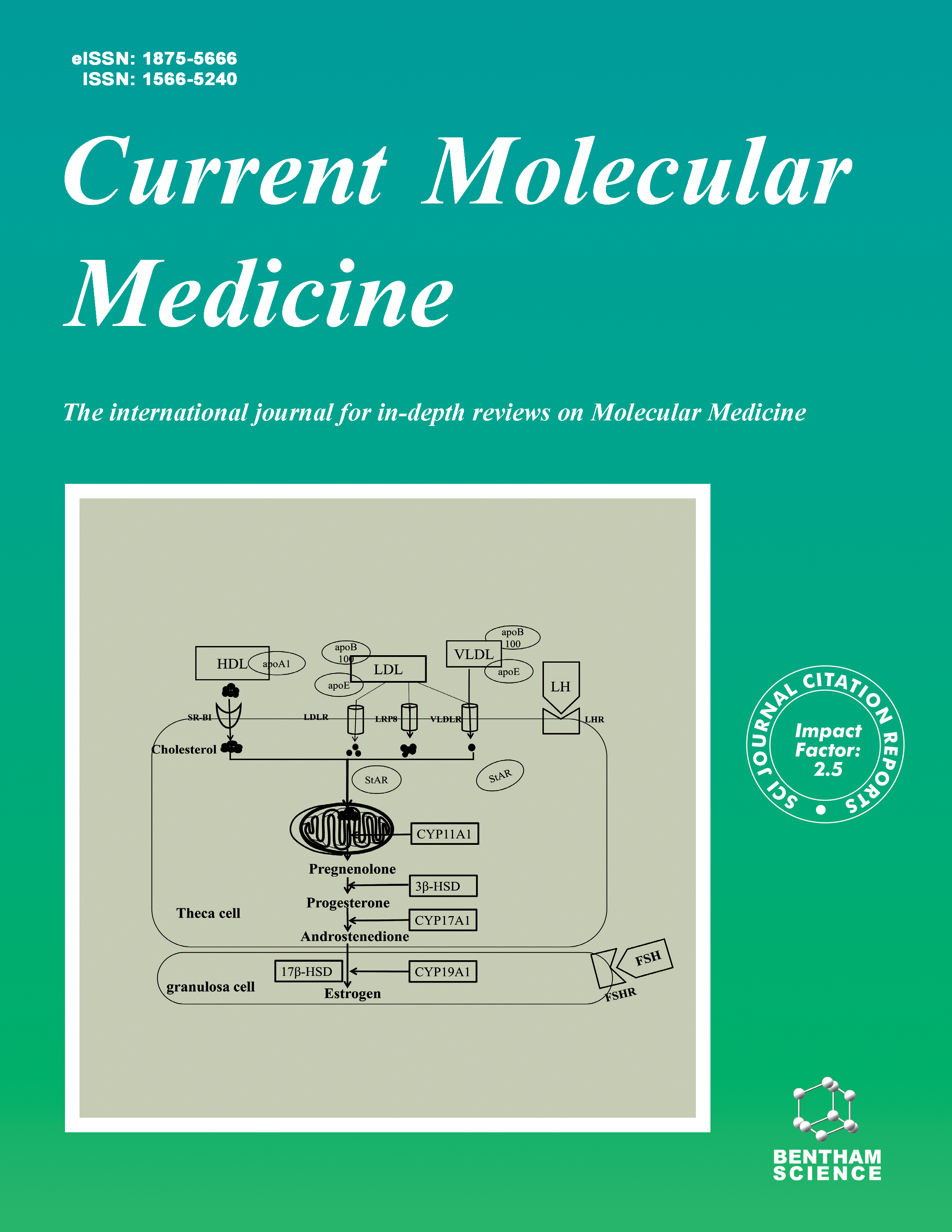
Current Molecular Medicine - Volume 25, Issue 3, 2025
Volume 25, Issue 3, 2025
-
-
The Emerging Role of LncRNA AWPPH in Multiple Cancers: A Review Study
More LessLong non-coding RNAs (lncRNAs) are transcribed RNA molecules longer than 200 nucleotides in length that have no protein-coding potential. They are able to react with DNA, RNA, and protein. Hence they involve in regulating gene expression at the epigenetic, transcriptional, post-transcriptional, and translational levels. LncRNAs have been proven to play an important role in human malignancies and prognostic outcomes. In this review, we will comprehensively and functionally discuss the role of a novel identified lncRNA, namely lncRNA WAPPH located on human chromosome 2q13, in various cancers. Increasing research studies have shown that lncRNA AWPPH is deregulated in different malignancies, including breast cancer, gastric cancer, colorectal cancer, ovarian cancer, bladder cancer, leukemia, and others. LncRNA WAPPH serves as an oncogene in tumorigenesis and the development of cancer. Moreover, lncRNA AWPPH is involved in numerous biological processes of solid and blood cancers. Taken together, based on our scrutiny analysis, lncRNA AWPPH can be regarded as a putative biomarker for diagnosis or therapeutic target in human malignancies.
-
-
-
Metabolic Derangement in Non-Alcoholic Fatty Liver Disease: Opportunities for Early Diagnostic and Prognostic Markers
More LessNon-alcoholic fatty liver disease is a globally prevalent disorder that can rapidly progress if not detected early. Currently, no accepted markers exist for early diagnosis and prognosis of NAFLD. This review describes derangement in major metabolic pathways of lipid, carbohydrate, and amino acids in NAFLD. It suggests that measuring levels of thrombospondin, TyG index, asymmetric dimethylarginine, LAL-A, GLP-1, FGF-21, and GSG index are potential markers for early diagnosis of NAFLD. A single marker may not indicate early NAFLD, and further large-scale studies on correlating levels of Thrombospondin-2, triglyceride-glucose index, and FGF-21 with NAFLD are warranted.
-
-
-
The Potential Therapeutic Applications of CRISPR/Cas9 in the Treatment of Gastrointestinal Cancers
More LessGastrointestinal (GI) cancer is one the most prevalent types of cancer. Despite current chemotherapy's success, patients with GI cancer continue to have a dismal outcome. The onset and progression of cancer are caused by alterations and the abnormal expression of several families of genes, like tumor-suppressor genes, oncogenes, and chemotherapy-resistant genes. The final purpose of tumor therapy is to inhibit cellular development by modifying mutations and editing the irregular expression of genes It has been reported that CDH1, TP53, KRAS, ARID1A, PTEN, and HLA-B are the commonly mutated genes in GI cancer. Gene editing has become one potential approach for cases with advanced or recurrent CRC, who are non-responsive to conventional treatments and a variety of driver mutations along with progression cause GI progression. CRISPR/Cas9 technique is a reliable tool to edit the genome and understand the functions of mutations driving GI cancer development. CRISPR/Cas9 can be applied to genome therapy for GI cancers, particularly with reference to molecular-targeted medicines and suppressors. Moreover, it can be used as a therapeutic approach by knocking in/out multiple genes. The use of CRISPR/ Cas9 gene editing method for GI cancer therapy has therefore resulted in some improvements. There are several research works on the role of CRISPR/Cas9 in cancer treatment that are summarized in the following separate sections. Here, the use of CRISPR/Cas9-based genome editing in GI and the use of CRISPR/Cas9 is discussed in terms of Targeting Chemotherapy Resistance-related Genes like; KRAS, TP53, PTEN, and ARID1A.
-
- Life Sciences, Biochemistry, Medicine, Research & Experimental, Biochemistry And Molecular Biology, Molecular Medicine
-
-
-
Current Perspectives on Attention-deficit Hyperactivity Disorder
More LessAuthors: Shaik Shafiullah and Suneela DhaneshwarAttention-deficit hyperactivity disorder (ADHD) is a neurobiological and neurodevelopmental disorder with an idiosyncratic genetic base. ADHD presents various characteristics, such as inattention, hyperactivity, and impulsivity. Over the period, ADHD leads to noticeable functional disability. A five- to ten-fold progressed risk of disorder development is observed in the populations with familial history of ADHD. The abnormal structure of the brain in ADHD results in altered neural mechanisms, such as cognition, attention, and memorial function. The mesolimbic, nigrostriatal, and mesocortical pathways in the brain get affected by the deterioration of the levels of dopamine. The hypothesis of dopamine in ADHD and its etiopathology suggests that detained attention and impaired arousal functions are due to reduced levels of do-pamine. The quickest way to improve strategical treatment is by clarifying the etiological aspects of ADHD and identifying the underlying mechanisms of pathophysiology, which will assist in exploring the biomarkers for better diagnosis. The implementation of life course theory is a very important research principle announced by Grand Challenges in Global Health Initiative (GCMHI). Long-term research is needed to define the progression of ADHD. Interdisciplinary collaborations promise a great future for research innovations in ADHD.
-
-
-
-
Implication of Thioredoxin 1 and Glutaredoxin 1 in H2O2-induced Phosphorylation of JNK and p38 MAP Kinases
More LessBackgroundAerobic organisms continuously generate small amounts of Reactive Oxygen Species (ROS), which are involved in the oxidation of sensitive cysteine residues in proteins, leading to the formation of disulfide bonds. Thioredoxin (Trx1) and Glutaredoxin (Grx1) represent key antioxidant enzymes reducing disulfide bonds.
ObjectiveIn this work, we have focused on the possible protective effect of Trx1 and Grx1 against oxidative stress-induced DNA damage and apoptosis-signaling, by studying the phosphorylation of MAP kinases.
MethodsTrx1 and Grx1 were overexpressed or silenced in cultured H1299 non-small cell lung cancer epithelial cells. We examined cell growth, DNA damage, and the phosphorylation status of MAP kinases following treatment with H2O2.
ResultsOverexpression of both Trx1 and Grx1 had a significant impact on the growth of H1299 cells and provided protection against H2O2-induced toxicity, as well as acute DNA single-strand breaks. Conversely, silencing of these proteins exacerbated DNA damage. Furthermore, overexpression of Trx1 and Grx1 inhibited the rapid phosphorylation of JNK (especially at 360 min of treatment, ****p=0.004 and **p=0.0033 respectively) and p38 MAP kinases (especially at 360 min of treatment, ****p<0.0001 and ***p=0.0008 respectively) during H2O2 exposure, while their silencing had the opposite effect (especially at 360 min of treatment, ****p<0.0001).
ConclusionThese results suggest that both Trx1 and Grx1 have protective roles against H2O2 induced toxicity, emphasizing their significance in mitigating oxidative stress-related cellular damage.
-
-
-
Exosomes Derived from Astragaloside IV-pretreated Endothelial Progenitor Cells (AS-IV-Exos) Alleviated Endothelial Oxidative Stress and Dysfunction via the miR-210/ Nox2/ROS Pathway
More LessAuthors: Wu Xiong, Xi Zhang, Xiao-Ling Zou, Sai Peng, Hua-Juan Lei, Xiang-Nan Liu, Lan Zhao and Zi-Xin HuangBackgroundChronic hyperglycemia in diabetes induces oxidative stress, leading to damage to the vascular system. In this study, we aimed to evaluate the effects and mechanisms of AS-IV-Exos in alleviating endothelial oxidative stress and dysfunction caused by high glucose (HG).
MethodsHistopathological changes were observed using HE staining, and CD31 expression was assessed through immunohistochemistry (IHC). Cell proliferation was evaluated through CCK8 and EDU assays. The levels of ROS, SOD, and GSH-Px in the skin tissues of each group were measured using ELISA. Cell adhesion, migration, and tube formation abilities were assessed using adhesion, Transwell, and tube formation experiments. ROS levels in HUVEC cells were measured using flow cytometry. The levels of miR-210 and Nox2 were determined through quantitative reverse transcription-polymerase chain reaction (qRT-PCR). The expression of Nox2, SOD, GSH-Px, CD63, and CD81 was confirmed using WB.
ResultsThe level of miR-210 was reduced in diabetes-induced skin damage, while the levels of Nox2 and ROS increased. Treatment with AS-IV increased the level of miR-210 in EPC-Exos. Compared to Exos, AS-IV-Exos significantly reduced the proliferation rate, adhesion number, migration speed, and tube-forming ability of HG-damaged HUVEC cells. AS-IV-Exos also significantly decreased the levels of SOD and GSH-Px in HG-treated HUVEC cells and reduced the levels of Nox2 and GSH-Px. However, ROS levels and Nox2 could reverse this effect.
ConclusionAS-IV-Exos effectively alleviated endothelial oxidative stress and dysfunction induced by HG through the miR-210/Nox2/ROS pathway.
-
-
-
Explore on the Mechanism of miRNA-146a/TAB1 in the Regulation of Cellular Apoptosis and Inflammation in Ulcerative Colitis Based on NF-κB Pathway
More LessAuthors: Xiaoying Xia, Qian Yang, Xue Han, Yulin Du, Shujun Guo, Mengqing Hua, Fang Fang, Zhigang Ma, Hua Ma, Hui Yuan, Wenjing Tian, Zebang Ding, Yanan Duan, Qi Huo and Yao LiObjectiveUlcerative colitis (UC) is a chronic non-specific inflammatory disease of the rectum and colon with unknown etiology. A growing number of evidence suggest that the pathogenesis of UC is related to excessive apoptosis and production of inflammatory cytokines. However, the functions and molecular mechanisms associated with UC remain unclear.
Materials and MethodsThe in vivo and in vitro models of UC were established in this study. MiRNA or gene expression was measured by qRT-PCR assay. ELISA, CCK-8, TUNEL, and flow cytometry assays were applied for analyzing cellular functions. The interactions between miR-146a and TAB1 were verified by luciferase reporter and miRNA pull-down assays.
ResultsMiR-146a was obviously increased in UC patients, DSS-induced colitis mice, and TNF-ɑ-induced YAMC cells, when compared to the corresponding controls. MiR-146a knockdown inhibited the inflammatory response and apoptosis in DSS-induced colitis mice and TNF-ɑ-induced YAMC cells. Mechanistically, we found that TAB1 was the target of miR-146a and miR-146a knockdown suppressed the activation of NF-κB pathway in UC. More importantly, TAB1 could overturn the inhibitory effect of antagomiR-146a on cell apoptosis and inflammation in UC.
ConclusionMiR-146a knockdown inhibited cell apoptosis and inflammation via targeting TAB1 and suppressing NF-κB pathway, suggesting that miR-146a may be a new therapeutic target for UC treatment.
-
-
-
Study on the Mechanism of Notch Pathway Mediates the Role of Lenvatinib-resistant Hepatocellular Carcinoma Based on Organoids
More LessAuthors: Weiqing Feng, Haixiong Zhang, Qing Yu, Hao Yin, Xiaowei Ou, Jie Yuan and Liang PengBackgroundThe emergence of treatment resistance has hindered the efficacy of targeted therapies used to treat patients with hepatocellular carcinoma (HCC).
ObjectiveThis study aimed to explore the mechanism of organoids constructed from lenvatinib-resistant HCC cells.
MethodsHep3B cell and human HCC organoids were cultured and identified using hematoxylin and eosin staining and Immunohistochemistry. Lenvatinib-sensitive/ resistant Hep3B cells were constructed using lenvatinib (0, 0.1, 1, and 10 μM) and lenvatinib (0, 1, 10, and 100 μM). qRT-PCR and flow cytometry were utilized to deter-mine HCC stem cell markers CD44, CD90, and CD133 expressions. Transcriptome sequencing was performed on organoids. Western blot evaluated Notch pathway-related proteins (NOTCH1 and Jagged) expressions. Furthermore, DAPT, an inhibitor of the Notch pathway, was used to investigate the effects of lenvatinib on resistance or stemness in organoids and human HCC tissues.
ResultsThe organoids were successfully cultivated. With the increase of lenvatinib concentration, sensitive cell organoids were markedly degraded and ATP activity was gradually decreased, while there was no significant change in ATP activity of resistant cell organoids. CD44 expressions were elevated after lenvatinib treatment compared with the control group. KEGG showed that lenvatinib treatment of organoids constructed from Hep3B cells mainly activated the Notch pathway. Compared with the control group, NOTCH1 and Jagged expressions elevated, and ATP activity decreased after lenvatinib treatment. However, ATP activity was notably decreased after DAPT treatment. Moreover, DAPT inhibited lenvatinib resistance and the increase in the expressions of CD44 caused by lenvatinib. Besides, 100 μM lenvatinib significantly inhibited the growth and ATP activity of human HCC organoids, and DAPT increased the inhibitory effect of lenvatinib.
ConclusionLenvatinib regulated resistance and stemness in organoids via the Notch pathway.
-
-
-
LKB1 Mutations Enhance Radiosensitivity in Non-Small Cell Lung Cancer Cells by Inducing G2/M Cell Cycle Phase Arrest
More LessAuthors: Yuanhu Yao, Xiangnan Qiu, Meng Chen, Zhaohui Qin, Xinjun Zhang and Wei ZhangBackgroundRadiosensitivity remains an important factor affecting the clinical outcome of radiotherapy for non-small cell lung cancer (NSCLC). Liver kinase B1 (LKB1) as a tumor suppressor, is one of the most commonly mutated genes in NSCLC. However, the role of LKB1 on radiosensitivity and the possible mechanism have not been elucidated in the NSCLC. In this study, we investigated the regulatory function of LKB1 in the radiosensitivity of NSCLC cells and its possible signaling pathways.
MethodsAfter regulating the expression of LKB1, cell proliferation was determined by Cell Counting Kit-8 (CCK-8) assay. The flow cytometry assay was used to analyse cell cycle distribution. Survival fraction and sensitization enhancement ratio (SER) were generated by clonogenic survival assay. Western blot analysis was used to assess expression levels of LKB1, p53, p21, γ-H2AX and p-Chk2.
ResultsOur study found that when the NSCLC cells were exposed to ionizing radiation, LKB1 could inhibit NSCLC cell proliferation by promoting DNA double strand break and inducing DNA repair. In addition, LKB1 could induce NSCLC cells G1 and G2/M phase arrest through up-regulating expression of p53 and p21 proteins.
ConclusionThis current study demonstrates that LKB1 enhances the radiosensitivity of NSCLC cells via inhibiting NSCLC cell proliferation and inducing G2/M phase arrest, and the mechanism of cell cycle arrest associated with signaling pathways of p53 and p21 probably.
-
Volumes & issues
-
Volume 25 (2025)
-
Volume 24 (2024)
-
Volume 23 (2023)
-
Volume 22 (2022)
-
Volume 21 (2021)
-
Volume 20 (2020)
-
Volume 19 (2019)
-
Volume 18 (2018)
-
Volume 17 (2017)
-
Volume 16 (2016)
-
Volume 15 (2015)
-
Volume 14 (2014)
-
Volume 13 (2013)
-
Volume 12 (2012)
-
Volume 11 (2011)
-
Volume 10 (2010)
-
Volume 9 (2009)
-
Volume 8 (2008)
-
Volume 7 (2007)
-
Volume 6 (2006)
-
Volume 5 (2005)
-
Volume 4 (2004)
-
Volume 3 (2003)
-
Volume 2 (2002)
-
Volume 1 (2001)
Most Read This Month


