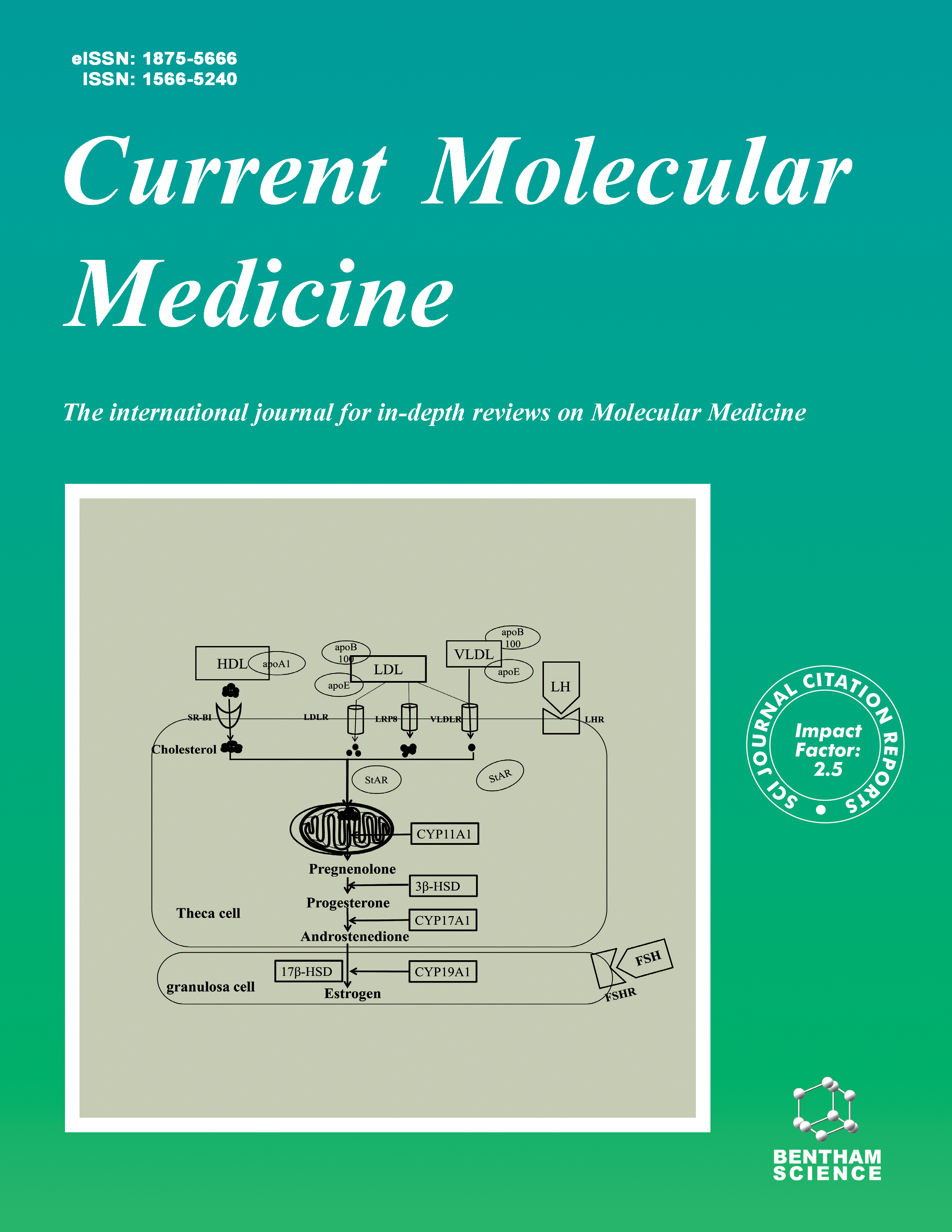
Current Molecular Medicine - Volume 24, Issue 3, 2024
Volume 24, Issue 3, 2024
-
-
Applications of Exosome Vesicles in Different Cancer Types as Biomarkers
More LessAuthors: Murat Ihlamur, Kübra Kelleci, Yağmur Zengin, M. Adil Allahverdiyev and Emrah Şefik AbamorOne of the biggest challenges in the fight against cancer is early detection. Early diagnosis is vital, but there are some barriers such as economic, cultural, and personal factors. Considering the disadvantages of radiological imaging techniques or serological analysis methods used in cancer diagnosis, such as being expensive, requiring expertise, and being time-consuming, there is a need to develop faster, more reliable, and cost-effective diagnostic methods for use in cancer diagnosis. Exosomes, which are responsible for intercellular communication with sizes ranging from 30-120 nm, are naturally produced biological nanoparticles. Thanks to the cargo contents they carry, they are a potential biomarker to be used in the diagnosis of cancer. Exosomes, defined as extracellular vesicles of endosomal origin, are effective in cancer growth, progression, metastasis, and drug resistance, and changes in microenvironmental conditions during tumor development change exosome secretion. Due to their high cellular activity, tumor cells produce much higher exosomes than healthy cells. Therefore, it is known that the number of exosomes in body fluids is significantly rich compared to other cells and can act as a stand-alone diagnostic biomarker. Cancer- derived exosomes have received great attention in recent years for the early detection of cancer and the evaluation of therapeutic response. In this article, the content, properties, and differences of exosomes detected in common types of cancer (lung, liver, pancreas, ovaries, breast, colorectal), which are the leading causes of cancer-related deaths, are reviewed. We also discuss the potential utility of exosome contents as a biomarker for early detection, which is known to be important in targeted cancer therapy.
-
-
-
Endoplasmic Reticulum as a Therapeutic Target in Cancer: Is there a Role for Flavonoids?
More LessFlavonoids are classified into subclasses of polyphenols, a multipurpose category of natural compounds which comprises secondary metabolites extracted from vascular plants and are plentiful in the human diet. Although the details of flavonoid mechanisms are still not realized correctly, they are generally regarded as antimicrobial, anti-fungal, anti-inflammatory, anti-oxidative; anti-mutagenic; anti-neoplastic; anti-aging; anti-diabetic, cardio-protective, etc. The anti-cancer properties of flavonoids are evident in functions such as prevention of proliferation, metastasis, invasion, inflammation and activation of cell death. Tumors growth and enlargement expose cells to acidosis, hypoxia, and lack of nutrients which result in endoplasmic reticulum (ER) stress; it triggers the unfolded protein response (UPR), which reclaims homeostasis or activates autophagy. Steady stimulation of ER stress can switch autophagy to apoptosis. The connection between ER stress and cancer, in association with UPR, has been explained. The signals provided by UPR can activate or inhibit anti-apoptotic or apoptotic pathways depending on the period and grade of ER stress. In this review, we will peruse the link between flavonoids and their impact on the endoplasmic reticulum in association with cancer therapy.
-
-
-
Neuroprotective Potential of Hesperidin as Therapeutic Agent in the Treatment of Brain Disorders: Preclinical Evidence-based Review
More LessAuthors: Keshav Bansal, Vanshita Singh, Sakshi Singh and Samiksha MishraNeurodegenerative disorders (NDs) are progressive morbidities that represent a serious health issue in the aging world population. There is a contemporary upsurge in worldwide interest in the area of traditional remedies and phytomedicines are widely accepted by researchers due to their health-promoted effects and fewer side effects. Hesperidin, a flavanone glycoside present in the peels of citrus fruits, possesses various biological activities including anti-inflammatory and antioxidant actions. In various preclinical studies, hesperidin has provided significant protective actions in a variety of brain disorders such as Alzheimer’s disease, epilepsy, Parkinson’s disease, multiple sclerosis, depression, neuropathic pain, etc. as well as their underlying mechanisms. The findings indicate that the neuroprotective effects of hesperidin are mediated by modulating antioxidant defence activities and neural growth factors, diminishing apoptotic and neuro-inflammatory pathways. This review focuses on the potential role of hesperidin in managing and treating diverse brain disorders.
-
-
-
Potential role of Nigella Sativa and its Constituent (Thymoquinone) in Ischemic Stroke
More LessAuthors: Shakiba Azami and Fatemeh ForouzanfarIschemic stroke is one of the major causes of global mortality, which puts great demands on health systems and social welfare. Ischemic stroke is a complex pathological process involving a series of mechanisms such as ROS accumulation, Ca2+ overload, inflammation, and apoptosis. The lack of effective and widely applicable pharmacological treatments for ischemic stroke patients has led scientists to find new treatments. The use of herbal medicine, as an alternative or complementary therapy, is increasing worldwide. For centuries, our ancestors had known the remedial nature of Nigella sativa (Family Ranunculaceae) and used it in various ways, either as medicine or as food. Nowadays, N. sativa is generally utilized as a therapeutic plant all over the world. Most of the therapeutic properties of this plant are attributed to the presence of thymoquinone which is the major biological component of the essential oil. The present review describes the pharmacotherapeutic potential of N. sativa in ischemic stroke that has been carried out by various researchers. Existing literature highlights the protective effects of N. sativa as well as thymoquinone in ischemia stroke via different mechanisms including anti-oxidative stress, anti-inflammation, anti-apoptosis, neuroprotective, and vascular protective effects. These properties make N. sativa and thymoquinone promising candidates for developing potential agents for the prevention and treatment of ischemic stroke.
-
-
-
Toxocara Canis Increases the Potential of Breast Cancer by Reducing the Expression of the P53 Protein
More LessIntroduction: Breast cancer is considered the most frequent type of cancer in women with high mortality worldwide, and most importantly, it is the second most common cancer. However, some breast cancer-related risk factors remain unknown. So, the current study was designed to evaluate the effect of Toxocara canis on the biomarkers correlated with proliferation, apoptosis, inflammation, and angiogenesis in 4T1 tumor-bearing mice infected with Toxocara canis for the first time. Methods: Mice were categorized into four groups: A) control, B) treated with 4T1+ Toxocara canis, C) treated with Toxocara canis, and D) treated with 4T1. The expression of Ki-67 and P53 was then evaluated by using the immunohistochemical technique. In addition, the levels of transforming growth factor-β, Interferon gamma-γ, Interleukin 10, tumor necrosis factor-α and vascular endothelial growth factor as well as anti- Toxocara canis IgG were determined using the enzyme-linked immunosorbent assay method. Results: The expression of Ki-67 was significantly increased in the 4T1+ Toxocara canis group than control and Toxocara canis groups (P < 0.001 and P < 0.001, respectively). Moreover, a significant decrease in P53 was found in the 4T1+ Toxocara canis group than in the control and Toxocara canis groups (P < 0.001 and P < 0.001, respectively). Also, the 4T1+ Toxocara canis group significantly reduced the expression of P53 more than 4T1 tumor-bearing mice (P = 0.005). In addition, the 4T1+ Toxocara canis group had an increasing tumor necrosis factor-α and vascular endothelial growth factor than controls (P = 0.004 and P = 0.002, respectively). Furthermore, a significant reduction in Interleukin 10 was found in the 4T1+ Toxocara canis group than in the control group (P = 0.004). Conclusion: Our findings showed that Toxocara canis could probably increase the potential of breast cancer by reducing P53 in 4T1 tumor-bearing mice infected with Toxocara canis more than other groups.
-
-
-
Albiflorin Alleviates Sepsis-induced Acute Liver Injury through mTOR/p70S6K Pathway
More LessAuthors: Yanan Liu, Lizhi Feng and Lan YaoBackground: Sepsis often induces hepatic dysfunction and inflammation, accounting for a significant increase in the incidence and mortality rates. To this end, albiflorin (AF) has garnered enormous interest due to its potent anti-inflammatory activity. However, the substantial effect of AF on sepsis-mediated acute liver injury (ALI), along with its potential mechanism of action, remains to be explored. Methods: An LPS-mediated primary hepatocyte injury cell model in vitro and a mouse model of CLP-mediated sepsis in vivo were initially built to explore the effect of AF on sepsis. Furthermore, the hepatocyte proliferation by CCK-8 assay in vitro and animal survival analyses in vivo for the survival time of mice were carried out to determine an appropriate concentration of AF. Then, flow cytometry, Western blot (WB), and TUNEL staining analyses were performed to investigate the effect of AF on the apoptosis of hepatocytes. Moreover, the expressions of various inflammatory factors by ELISA and RT-qPCR analyses and oxidative stress by ROS, MDA, and SOD assays were determined. Finally, the potential mechanism of AF alleviating the sepsis-mediated ALI via the mTOR/p70S6K pathway was explored through WB analysis. Results: AF treatment showed a significant increase in the viability of LPS-inhibited mouse primary hepatocytes cells. Moreover, the animal survival analyses of the CLP model mice group indicated a shorter survival time than the CLP+AF group. AF-treated groups showed significantly decreased hepatocyte apoptosis, inflammatory factors, and oxidative stress. Finally, AF exerted an effect by suppressing the mTOR/p70S6K pathway. Conclusion: In summary, these findings demonstrated that AF could effectively alleviate sepsis-mediated ALI via the mTOR/p70S6K signaling pathway.
-
-
-
Baicalein Alleviates Arsenic-induced Oxidative Stress through Activation of the Keap1/Nrf2 Signalling Pathway in Normal Human Liver Cells
More LessAuthors: Qi Wang and Aihua ZhangBackground: Oxidative stress is a key mechanism underlying arsenicinduced liver injury, the Kelch-like epichlorohydrin-related protein 1 (Keap1)/nuclear factor E2 related factor 2 (Nrf2) pathway is the main regulatory pathway involved in antioxidant protein and phase II detoxification enzyme expression. The aim of the present study was to investigate the role and mechanism of baicalein in the alleviation of arsenic-induced oxidative stress in normal human liver cells. Methods: Normal human liver cells (MIHA cells) were treated with NaAsO2 (0, 5, 10, 20 μM) to observe the effect of different doses of NaAsO2 on MIHA cells. In addition, the cells were treated with DMSO (0.1%), NaAsO2 (20 μM), or a combination of NaAsO2 (20 μM) and Baicalein (25, 50 or 100 μM) for 24 h to observe the antagonistic effect of Baicalein on NaAsO2. Cell viability was determined using a Cell Counting Kit- 8 (CCK-8 kit). The intervention doses of baicalein in subsequent experiments were determined to be 25, 50 and 100μM. The intracellular content of reactive oxygen species (ROS) was assessed using a 2′,7′-dichlorodihydrofluorescein diacetate (DCFHDA) probe kit. The malonaldehyde (MDA), Cu-Zn superoxide dismutase (Cu-Zn SOD) and glutathione peroxidase (GSH-Px) activities were determined by a test kit. The expression levels of key genes and proteins were determined by real-time fluorescence quantitative polymerase chain reaction (qPCR) and Western blotting. Results: Baicalein upregulated the protein expression levels of phosphorylated Nrf2 (p-Nrf2) and nuclear Nrf2, inhibited the downregulation of Nrf2 target genes induced by arsenic, and decreased the production of ROS and MDA. These results demonstrate that baicalein promotes Nrf2 nuclear translocation by upregulating p-Nrf2 and inhibiting the downregulation of Nrf2 target genes in arsenic-treated MIHA cells, thereby enhancing the antioxidant capacity of cells and reducing oxidative stress. Conclusion: Baicalein alleviated arsenic-induced oxidative stress through activation of the Keap1/Nrf2 signalling pathway in normal human liver cells.
-
-
-
Elevation of LEM Domain Containing 1 Predicts Poor Prognosis of NSCLC Patients and Triggers Malignant Stemness and Invasion of NSCLC Cells by Stimulating PI3K/AKT Pathway
More LessBackground: Non-small cell lung cancer (NSCLC) is a leading cause of cancer-related death globally. LEM domain containing 1 (LEMD1) function has been identified in several cancers but not in NSCLC. Objective: This study aimed to investigate the LEMD1 function in NSCLC. Methods: NSCLC tissues were obtained from 66 patients, and LEMD1 expressions were measured using quantitative real-time PCR, immunohistochemical assay, and Western blot. Overall survival of NSCLC patients was estimated by the Kaplan-Meier method. Meanwhile, LEMD1 function and mechanism were assessed using Cell Counting Kit-8, 5-Ethynyl-2′-deoxyuridine analysis, Transwell, Sphere formation assay, and flow cytometry. Furthermore, LEMD1 function in vivo was evaluated by establishing a xenograft tumor model, hematoxylin-eosin staining, and immunohistochemical assay. Results: LEMD1 was highly expressed in NSCLC tissues and was interrelated to tumor differentiation, TNM stage, and lymph node metastasis of patients. Overall survival of NSCLC patients with high LEMD1 was found to be lower than that of patients with low LEMD1. Functionally, interference with LEMD1 restrained NSCLC cell proliferation, invasion, and stemness characteristics. Mechanistically, LEMD1 facilitated the malignant phenotype of NSCLC, and 740 Y-P reversed this impact, prompting that LEMD1 aggravated NSCLC by activating PI3K/AKT pathway. Furthermore, LEMD1 knockdown hindered NSCLC proliferation in vivo. Conclusion: LEMD1 accelerated NSCLC cell proliferation, invasion, and stemness characteristics via activating PI3K/AKT pathway.
-
-
-
MT1G Regulates c-MYC/P53 Signal to Inhibit Proliferation, Invasion and Migration and Promote Apoptosis in Colon Cancer Cells
More LessAuthors: Jie Li, Qiaozhen Hu, Zhongyan Li, Kaiyu Feng and Kangbao LiIntroduction: Colon cancer is a common and malignant cancer featuring high morbidity and poor prognosis. Aims: This study was performed to explore the regulatory role of MT1G in colon cancer as well as its unconcealed molecular mechanism. Methods: The expressions of MT1G, c-MYC, and p53 were assessed with the application of RT-qPCR and western blot. The impacts of MT1G overexpression on the proliferative ability of HCT116 and LoVo cells were measured by CCK-8 and BrdU incorporation assays. Additionally, transwell wound healing, and flow cytometry assays were employed to evaluate the invasive and migrative capacities as well as the apoptosis level of HCT116 and LoVo cells. Moreover, the activity of the P53 promoter region was assessed with the help of a luciferase reporter assay. Results: It was found that the expressions of MT1G at both mRNA and protein levels were greatly decreased in human colon cancer cell lines, particularly in HCT116 and LoVo cell lines. After transfection, it was discovered that the MT1G overexpression suppressed the proliferation, migration and invasion but promoted the apoptosis of HCT116 and LoVo cells, which were then partially reversed after overexpressing c-MYC. Additionally, MT1G overexpression reduced c-MYC expression but enhanced the p53 expression, revealing that the MT1G overexpression could regulate c-MYC/P53 signal. Elsewhere, it was also shown that c-MYC overexpression suppressed the regulatory effects of MT1G on P53. Conclusion: To conclude, MT1G was verified to regulate c-MYC/P53 signal to repress the proliferation, migration and invasion but promote the apoptosis of colon cancer cells, which might offer a novel targeted-therapy for the improvement of colon cancer.
-
-
-
Pre-clinical Efficacy and Safety Pharmacology of PEGylated Recombinant Human Endostatin
More LessAuthors: Lifang Guo, Linbin Hua, Bin Hu and Jing WangIntroduction: This study aimed to outline the pre-clinical efficacy and safety pharmacology of PEGylated recombinant human endostatin (M2ES) according to the requirements of new drug application. Methods: The purity of M2ES was evaluated by using silver staining. Transwell migration assay was applied to detect the bioactivity of M2ES in vitro. The antitumor efficacy of M2ES was evaluated in an athymic nude mouse xenograft model of pancreatic cancer (Panc-1) and gastric cancer (MNK45). BALB/C mice were treated with different doses of M2ES (6, 12 and 24 mg/kg) intravenously, both autonomic activity and cooperative sleep were monitored before and after drug administration. Results: The apparent molecular weight of M2ES was about 50 kDa, and the purity was greater than 98%. Compared with the control group, M2ES significantly inhibits human micro-vascular endothelial cells (HMECs) migration in vitro. Notably, weekly administration of M2ES showed a significant antitumor efficacy when compared with the control group. Treatment of M2ES (24mg/kg or below) showed no obvious effect on both autonomic activity and hypnosis. Conclusion: On the basis of the pre-clinical efficacy and safety pharmacology data of M2ES, M2ES can be authorized to carry out further clinical studies.
-
Volumes & issues
-
Volume 25 (2025)
-
Volume 24 (2024)
-
Volume 23 (2023)
-
Volume 22 (2022)
-
Volume 21 (2021)
-
Volume 20 (2020)
-
Volume 19 (2019)
-
Volume 18 (2018)
-
Volume 17 (2017)
-
Volume 16 (2016)
-
Volume 15 (2015)
-
Volume 14 (2014)
-
Volume 13 (2013)
-
Volume 12 (2012)
-
Volume 11 (2011)
-
Volume 10 (2010)
-
Volume 9 (2009)
-
Volume 8 (2008)
-
Volume 7 (2007)
-
Volume 6 (2006)
-
Volume 5 (2005)
-
Volume 4 (2004)
-
Volume 3 (2003)
-
Volume 2 (2002)
-
Volume 1 (2001)
Most Read This Month


