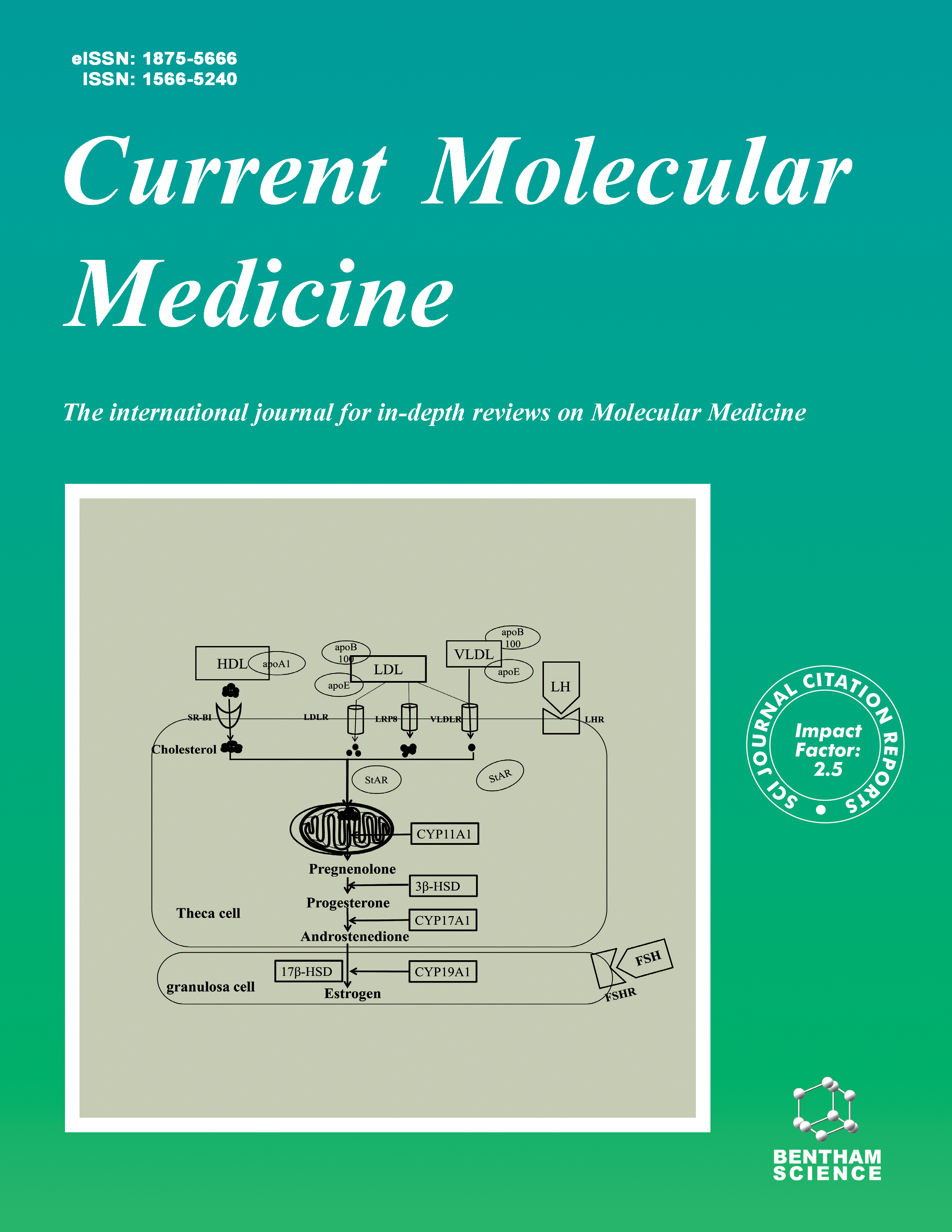
Current Molecular Medicine - Volume 24, Issue 2, 2024
Volume 24, Issue 2, 2024
-
-
Circular RNAs: Emerging Modulators in the Pathophysiology of Polycystic Ovary Syndrome and their Clinical Implications
More LessAuthors: Sahar Mazloomi, Vahide Mousavi, Esmat Aghadavod and Alireza MafiPolycystic ovary syndrome (PCOS) is a prevalent endocrine/metabolic disorder in women of reproductive age. PCOS is characterized by hyperandrogenism, polycystic ovary morphology, and ovulatory dysfunction/anovulation. It involves multiple effects in patients, including granulosa/theca cell hyperplasia, menstrual disturbances, infertility, acne, obesity, insulin resistance, and cardiovascular disorders.Biochemical analyses and the results of RNA sequencing studies in recent years have shown a type of non-coding RNAs as a splicing product known as circular RNAs (circRNAs). Several biological functions have been identified in relation to circRNAs, including a role in miRNA sponge, protein sequestration, increased parental gene expression, and translation leading to polypeptides. These circular molecules are more plentiful and specialized than other types of RNAs. For this reason, they are referred to as potential biomarkers in different diseases. Evidence suggests that circRNAs may have regulatory potentials through different signaling pathways, such as the miRNA network.Probably most experts in the field of obstetricians are not aware of circRNAs as a useful biomarker. Therefore, this review focused on the researches that have been done on the involvement of circRNAs in PCOS and summarized recent supportive evidence, and evaluated the circRNA association and mechanisms involved in PCOS.
-
-
-
The Effect of Bacterial Composition Shifts in the Oral Microbiota on Alzheimer's Disease
More LessAlzheimer's disease (AD), a neurological disorder, despite significant advances in medical science, has not yet been definitively cured, and the exact causes of the disease remain unclear. Due to the importance of AD in the clinic, large expenses are spent annually to deal with this neurological disorder, and neurologists warn of an increase in this disease in elderly in the near future. It has been believed that microbiota dysbiosis leads to Alzheimer's as a multi-step disease. In this regard, the presence of footprints of perturbations in the oral microbiome and the predominance of pathogenic bacteria and their effect on the nervous system, especially AD, is a very interesting topic that has been considered by researchers in the last decade. Some studies have looked at the mechanisms by which oral microbiota cause AD. However, many aspects of this interaction are still unclear as to how oral microbiota composition can contribute to this disease. Understanding this interaction requires extensive collaboration by interdisciplinary researchers to explore all aspects of the issue. In order to reveal the link between the composition of the oral microbiota and this disease, researchers from various domains have sought to explain the mechanisms of shift in oral microbiota in AD in this review.
-
-
-
Autophagy as an Anti-senescent in Aging Neurocytes
More LessNeuron homeostasis is crucial for the organism, and its maintenance is multifactorial, including autophagy. The turnover of aberrant intracellular components is a fundamental pathogenetic mechanism for cell aging. Autophagy is involved in the acceleration of the neurocyte aging process and the modification of cell longevity. Neurocyte aging is a process of loss of cell identity through cellular and subcellular changes that include molecular loss of epigenetics, transcriptomic, proteomic, and autophagy dysfunction. Autophagy dysfunction is the hallmark of neurocyte aging. Cell aging is the credential feature of neurodegenerative diseases. Pathophysiologically, aged neurocytes are characterized by dysregulated autophagy and subsequently neurocyte metabolic stress, resulting in accelerated neurocyte aging. In particular, chaperone- mediated autophagy perturbation results in upregulated expression of aging and apoptosis genes. Aged neurocytes are also characterized by the down-regulation of autophagy-related genes, such as ATG5-ATG12, LC3-II / LC3-I ratio, Beclin-1, and p62. Slowing aging through autophagy targeting is sufficient to improve prognosis in neurodegenerative diseases. Three primary anti-senescent molecules are involved in the aging process: mTOR, AMPK, and Sirtuins. Autophagy therapeutic effects can be applied to reverse and slow aging. This article discusses current advances in the role of autophagy in neurocyte homeostasis, aging, and potential therapeutic strategies to reduce aging and increase cell longevity.
-
-
-
CRISPR-Cas9: A Potent Gene-editing Tool for the Treatment of Cancer
More LessThe prokaryotic adaptive immune system has clustered regularly interspaced short palindromic repeat. CRISPR-associated protein (CRISPR-Cas) genome editing systems have been harnessed. A robust programmed technique for efficient and accurate genome editing and gene targeting has been developed. Engineered cell therapy, in vivo gene therapy, animal modeling, and cancer diagnosis and treatment are all possible applications of this ground-breaking approach. Multiple genetic and epigenetic changes in cancer cells induce malignant cell growth and provide chemoresistance. The capacity to repair or ablate such mutations has enormous potential in the fight against cancer. The CRISPR-Cas9 genome editing method has recently become popular in cancer treatment research due to its excellent efficiency and accuracy. The preceding study has shown therapeutic potential in expanding our anticancer treatments by using CRISPR-Cas9 to directly target cancer cell genomic DNA in cellular and animal cancer models. In addition, CRISPR-Cas9 can combat oncogenic infections and test anticancer medicines. It may design immune cells and oncolytic viruses for cancer immunotherapeutic applications. In this review, these preclinical CRISPRCas9- based cancer therapeutic techniques are summarised, along with the hurdles and advancements in converting therapeutic CRISPR-Cas9 into clinical use. It will increase their applicability in cancer research.
-
-
-
Oxidative versus Reductive Stress in Breast Cancer Development and Cellular Mechanism of Alleviation: A Current Perspective with Anti-breast Cancer Drug Resistance
More LessAuthors: Suman K. Ray, Erukkambattu Jayashankar, Ashwin Kotnis and Sukhes MukherjeeRedox homeostasis is essential for keeping our bodies healthy, but it also helps breast cancer cells grow, stay alive, and resist treatment. Changes in the redox balance and problems with redox signaling can make breast cancer cells grow and spread and make them resistant to chemotherapy and radiation therapy. Reactive oxygen species/reactive nitrogen species (ROS/RNS) generation and the oxidant defense system are out of equilibrium, which causes oxidative stress. Many studies have shown that oxidative stress can affect the start and spread of cancer by interfering with redox (reduction-oxidation) signaling and damaging molecules. The oxidation of invariant cysteine residues in FNIP1 is reversed by reductive stress, which is brought on by protracted antioxidant signaling or mitochondrial inactivity. This permits CUL2FEM1B to recognize its intended target. After the proteasome breaks down FNIP1, mitochondrial function is restored to keep redox balance and cell integrity. Reductive stress is caused by unchecked amplification of antioxidant signaling, and changes in metabolic pathways are a big part of breast tumors' growth. Also, redox reactions make pathways like PI3K, PKC, and protein kinases of the MAPK cascade work better. Kinases and phosphatases control the phosphorylation status of transcription factors like APE1/Ref-1, HIF-1, AP-1, Nrf2, NF-B, p53, FOXO, STAT, and - catenin. Also, how well anti-breast cancer drugs, especially those that cause cytotoxicity by making ROS, treat patients depends on how well the elements that support a cell's redox environment work together. Even though chemotherapy aims to kill cancer cells, which it does by making ROS, this can lead to drug resistance in the long run. The development of novel therapeutic approaches for treating breast cancer will be facilitated by a better understanding of the reductive stress and metabolic pathways in tumor microenvironments.
-
-
-
Hsa_circ_0004662 Accelerates the Progression of Osteoarthritis via the microRNA-424-5p/VEGFA Axis
More LessAuthors: Wei Xie, Luoyong Jiang, Xiaoyang Huang, Wei You and Wei SunObjective: Circular RNAs (circRNAs) have been extensively implicated in osteoarthritis (OA) progression. Therefore, this study explores the impact of hsa_circ_0004662 on OA progression and the related molecular mechanism.Methods: Human articular chondrocyte injury was induced by IL-1β to construct the OA model in vitro. Hsa_circ_0004662 and microRNA (miR)-424-5p expression in chondrocytes was evaluated with qRT-PCR. Vascular endothelial growth factors A (VEGFA) expression was examined with qRT-PCR and western blot after hsa_circ_0004662 knockdown or miR-424-5p overexpression in chondrocytes. Subsequent to loss- and gain-of-function assays in IL-1β-induced chondrocytes, the proliferation and apoptosis of chondrocytes were assessed with CCK-8 assay and flow cytometry, respectively. The expression of MMP13, Aggrecan, and apoptosis-related proteins Bax and Bcl-2 was measured with western blot. The binding of miR-424-5p to hsa_circ_0004662 and VEGFA was assessed with a dual-luciferase reporter gene assay.Results: Hsa_circ_0004662 was up-regulated, but miR-424-5p was down-regulated in IL-1β-induced chondrocytes. Mechanistically, both hsa_circ_0004662 and VEGFA bound to miR-424-5p, and hsa_circ_0004662 enhanced VEGFA expression by downregulating miR-424-5p. Hsa_circ_0004662 knockdown elevated cell proliferation, decreased apoptosis and MMP13 and Bax expression, and increased Aggrecan and Bcl- 2 expression in IL-1β-induced chondrocytes, which was counteracted by further miR- 424-5p down-regulation or VEGFA overexpression.Conclusion: Hsa_circ_0004662 facilitates OA progression via the miR-424-5p/ VEGFA axis.
-
-
-
The Diagnostic and Prognostic Value of Plasma Circulating CircNUP98 for Nasopharyngeal Carcinoma
More LessAuthors: Bin Zhang, Bohuai Xu, Lujie Yu, Yingying Pei and Yong HeBackground: Our preliminary sequencing analysis revealed increased expression levels of circNUP98 in nasopharyngeal carcinoma (NPC). This study was therefore carried out to explore the role of circNUP98 in NPC.Methods: The present study enrolled 56 patients with NPC, 44 patients with cervical lymphadenitis (CL), 50 patients with nose bleeding (NB), 50 patients with chronic sinusitis (CS), 50 patients with lymph node tuberculosis (LNT), and 50 healthy controls (Control). Plasma samples were obtained from all patients and the controls. In addition, NPC and paired non-tumor tissue samples were collected from the 56 NPC patients. The expression of circNUP98 in both tissue and plasma samples was determined by RT-qPCR. The 56 NPC patients were followed up for 5 years to analyze the associations between plasma expression of circNUP98 and the survival of patients. The diagnostic value of circNUP98 for NPC was analyzed through ROC curve analysis.Results: The plasma expression levels of circNUP98 were only increased in NPC, but not in CL, NB, CS and LNT groups compared to that in the Control group. In addition, increased expression levels of circNUP98 were observed in NPC tissues compared to that in non-tumor tissues. Plasma circNUP98 was closely correlated with circNUP98 in NPC tissues, but not circNUP98 in non-tumor tissues. With plasma circNUP98 as a biomarker, NPC patients were separated from CL, NB, CS, LNT, and the Control groups. The Plasma expression of circNUP98 was found to be positively correlated with the poor survival of patients. Moreover, plasma circNUP98 was only closely associated with tumor metastasis, but not tumor size.Conclusion: The expression of circNUP98 in plasma may be used to diagnose NPC and predict patients' survival.
-
-
-
Time- and Concentration-Dependent Stimulation of Oxidative Stress in Chondrocytes by Intracellular Soluble Urate
More LessAuthors: Bingqing Zhang, Hong Di, Yun Zhang, Xinxin Han, Yue Yin, Yingdong Han, Yu Cao and Xuejun ZengBackground: Gout could result in irreversible bone erosion, and chondrocyte might be involved in the process. Increased soluble urate is the early stage of gout and is strongly oxidative.Objective: To explore the effect of intracellular urate on the oxidative status of chondrocytes.Methods: A chondrocyte model was used. Serial concentrations of exogenous urate were incubated with chondrocytes for increasing amounts of time. Reactive oxygen species (ROS), oxidant, and anti-oxidant molecules were measured with biochemical assays, rt-PCR, and western blot. A urate transport inhibitor and oxidative inhibitors were used to confirm the effect of exogenous urate.Results: All concentrations of exogenous urate stimulated the production of ROS in a time- and concentration-dependent manner, as well as oxidant molecules, including hydrogen peroxide (H2O2), nicotinamide adenine dinucleotide phosphate (NADPH) oxidase, nitric oxide (NO) inducible nitric oxide synthase (iNOS), and these effects, could be inhibited by oxidant inhibitors. However, anti-oxidant molecules, including acidic leucine-rich nuclear phosphoprotein-32A (ANP32A), ataxia-telangiectasia mutated (ATM), heme oxygenase-1 (HO-1), and the transcription factor nuclear factor erythroid 2 (NF-E2)-related (Nrf2), was decreased by high concentrations of exogenous urate after prolonged incubation, but not by low to medium concentrations of exogenous urate. By inhibiting soluble urate trafficking, benzbromarone significantly suppressed the effect of urate stimulus on the oxidant and anti-oxidant molecules.Conclusion: Intracellular soluble urate could regulate chondrocyte redox balance in a time and concentration-dependent manner, and would be a target for regulating and protecting chondrocyte function in the early gout stage.
-
-
-
Expression of Matrix Metalloproteinases in Human Cystic Echinococcosis
More LessBackground: Cystic echinococcosis (CE) is a zoonotic disease caused by the Echinococcus granulosus senso lato (E. granulosus s.l.) larval stages. Parasitederived products have been shown to regulate host matrix metalloproteinases (MMPs), contributing to CE pathogenesis and progressive liver fibrosis in intermediate hosts. The current study aimed to investigate the potential role of MMP1, 7, 8, and 13 in E. granulosus s.l-induced liver fibrosis.Methods: Thirty CE patients with active, transitional, or inactive hydatid cysts were enrolled in this study to determine the inductive effects of E. granulosus on the expression of MMP-1, MMP-7, MMP-8, and MMP-13 in healthy liver tissue and fibrotic liver tissue using qRT-PCR.Results: According to the WHO-IWGE classification, patients with functional cysts (CE1 and CE2) had the highest percentage (46.6%). MMP-1, MMP-7, MMP-8, and MMP-13 expression levels were significantly higher in fibrotic liver than in normal liver tissue. MMP-13 and MMP-1 had the highest and lowest expression levels among MMPs. Compared to the normal group, the fold change for MMP-13 in the fibrotic group was greater than 12 and had the highest AUC value (AUC= 0.8283).Conclusion: Our findings suggest that E. granulosus-derived products might be involved in regulating host MMPs. Thus, MMPs may be considered potential biomarkers for predicting CE prognosis. Because of the non-normal distribution of our patients' CE types, further research, particularly on circulation MMPs, is needed to confirm the potential role of MMPs in CE pathogenesis and to follow up on CE patients.
-
-
-
Astragaloside IV (ASIV) Mediates Endothelial Progenitor Cell (EPC) Exosomal LINC01963 to Inhibit Pyroptosis and Oxidative Stress in High Glucose-impaired Endothelial Cells
More LessAuthors: Wu Xiong, Xi Zhang, Jian-da Zhou, Mei-xin Tan, Yu Liu, Yu Yan, Hua-Juan Lei, Jia-rui Peng, Wei Liu and Pei TanBackground: Hyperglycemia is widespread in the world's population, increasing the risk of many diseases. This study aimed to explore the regulatory effect and mechanism of astragaloside IV (ASIV)-mediated endothelial progenitor cells (EPCs) exosomal LINC01963 in endothelial cells (HUVECs) impaired by high glucose.Methods: Morphologies of exosomes were observed by light microscope and electron microscope. Immunofluorescence was used to identify EPCs and detect the expressions of caspase-1. LINC01963 was detected by quantitative reverse transcription PCR. NLRP3, ASC, and caspase-3 were detected by Western Blot. Nanoparticle tracking analysis was carried out to analyze the exosome diameter. High-throughput sequencing was applied to screen target lncRNAs. The proliferation of endothelial cells was measured by cell counting kit-8 assay. The apoptosis level of HUVECs was detected by flow cytometry and TdT-mediated dUTP Nick-End labeling. The levels of IL- 1β, IL-18, ROS, SOD, MDA, and LDH were measured by enzyme-linked immunosorbent assay.Results: ASIV could promote the secretion of the EPC exosome. LINC01963 was obtained by high-throughput sequencing. It was observed that high glucose could inhibit the proliferation, reduce the level of SOD, the expression of NLRP3, ASC, and caspase- 1, increase the levels of IL-1β, IL-18, ROS, MDA, and LDH, and promote apoptosis of HUVECs. Whereas LINC01963 could inhibit the apoptosis of HUVECs, the increase the expression of NLRP3, ASC, and caspase-1, and decrease the levels of IL-1β, IL-18, ROS, MDA, and LDH.Conclusion: EPCs exosomal LINC01963 play an inhibitory role in high glucoseinduced pyroptosis and oxidative stress of HUVECs. This study provides new ideas and directions for treating hyperglycemia and researching exosomal lncRNAs.
-
-
-
Intracellular Ellagic Acid Derived from Goat Urine DMSO Fraction (GUDF) Predicted as an Inhibitor of c-Raf Kinase
More LessBackground: Dietary chemicals and their gut-metabolized products are explored for their anti-proliferative and pro-cell death effects. Dietary and metabolized chemicals are different from ruminants such as goats over humans.Methods: Loss of cell viability and induction of death due to goat urine DMSO fraction (GUDF) derived chemicals were assessed by routine in vitro assays upon MCF-7 breast cancer cells. Intracellular metabolite profiling of MCF-7 cells treated with goat urine DMSO fraction (GUDF) was performed using an in-house designed vertical tube gel electrophoresis (VTGE) assisted methodology, followed by LC-HRMS. Next, identified intracellular dietary chemicals such as ellagic acid were evaluated for their inhibitory effects against transducers of the c-Raf signaling pathway employing molecular docking and molecular dynamics (MD) simulation.Results: GUDF treatment upon MCF-7 cells displayed significant loss of cell viability and induction of cell death. A set of dietary and metabolized chemicals in the intracellular compartment of MCF-7 cells, such as ellagic acid, 2-hydroxymyristic acid, artelinic acid, 10-amino-decanoic acid, nervonic acid, 2,4-dimethyl-2-eicosenoic acid, 2,3,4'- Trihydroxy,4-Methoxybenzophenone and 9-amino-nonanoic acid were identified. Among intracellular dietary chemicals, ellagic acid displayed a strong inhibitory affinity (-8.7 kcal/mol) against c-Raf kinase. The inhibitory potential of ellagic acid was found to be significantly comparable with a known c-Raf kinase inhibitor sorafenib with overlapping inhibitory site residues (ARG450, GLU425, TRP423, VA403).Conclusion: Intracellular dietary-derived chemicals such as ellagic acid are suggested for the induction of cell death in MCF-7 cells. Ellagic acid is predicted as an inhibitor of c-Raf kinase and could be explored as an anti-cancer drug.
-
Volumes & issues
-
Volume 25 (2025)
-
Volume 24 (2024)
-
Volume 23 (2023)
-
Volume 22 (2022)
-
Volume 21 (2021)
-
Volume 20 (2020)
-
Volume 19 (2019)
-
Volume 18 (2018)
-
Volume 17 (2017)
-
Volume 16 (2016)
-
Volume 15 (2015)
-
Volume 14 (2014)
-
Volume 13 (2013)
-
Volume 12 (2012)
-
Volume 11 (2011)
-
Volume 10 (2010)
-
Volume 9 (2009)
-
Volume 8 (2008)
-
Volume 7 (2007)
-
Volume 6 (2006)
-
Volume 5 (2005)
-
Volume 4 (2004)
-
Volume 3 (2003)
-
Volume 2 (2002)
-
Volume 1 (2001)
Most Read This Month


