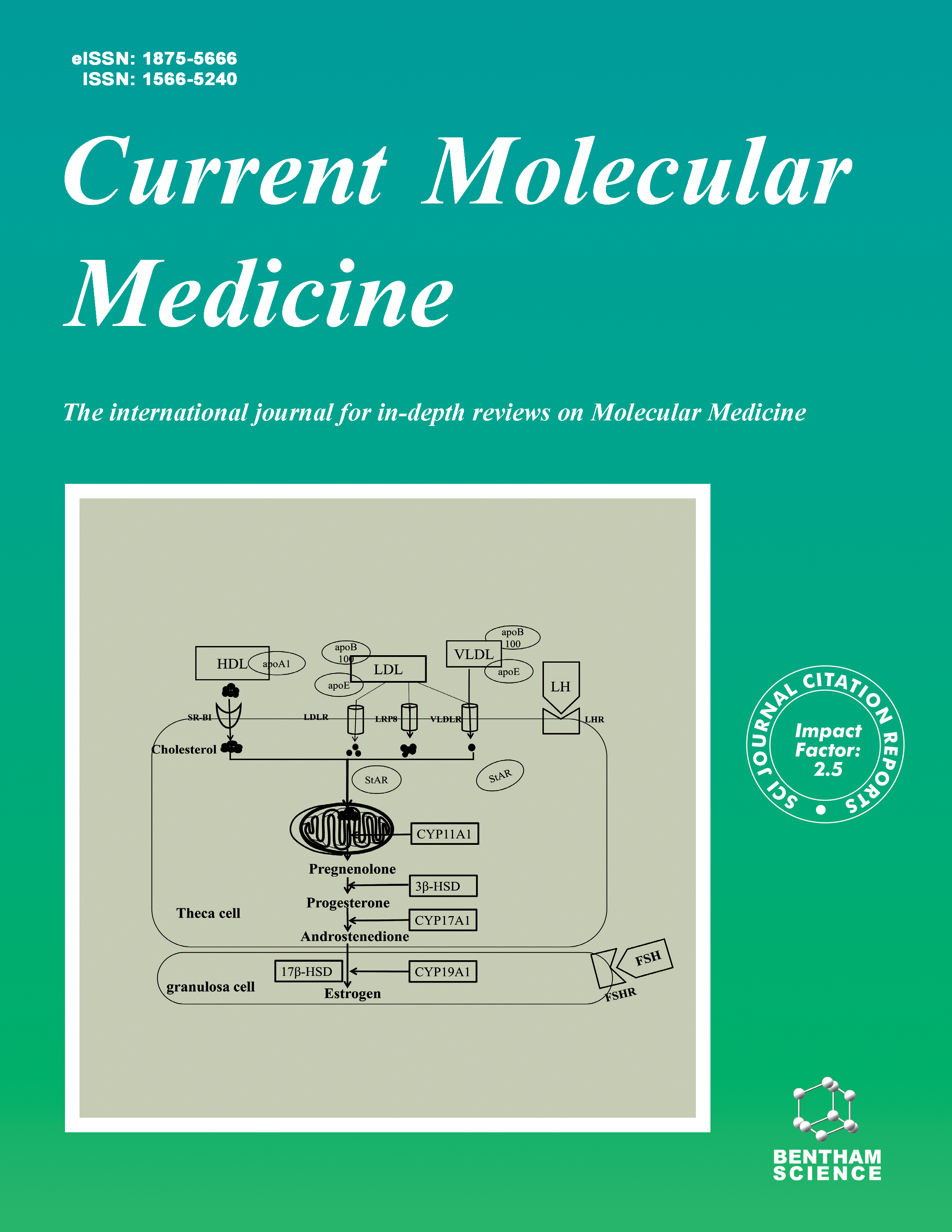
Current Molecular Medicine - Volume 20, Issue 7, 2020
Volume 20, Issue 7, 2020
-
-
Alzheimer’s Disease: A Contextual Link with Nitric Oxide Synthase
More LessAuthors: Harikesh Dubey, Kavita Gulati and Arunabha RayNitric oxide (NO) is a gasotransmitter with pleiotropic effects which has made a great impact on biology and medicine. A multidimensional neuromodulatory role of NO has been shown in the brain with specific reference to neurodegenerative disorders like Alzheimer’s disease (AD) and cognitive dysfunction. It has been found that NO/cGMP signalling pathway has an important role in learning and memory. Initially, it was considered that indirectly NO exerted neurotoxicity in AD via glutamatergic excitotoxicity. However, considering the early development of cognitive functions involved in the learning memory process including long term potentiation and synaptic plasticity, NO has a crucial role. Increasing evidence uncovered the above facts that isoforms of NOS viz endothelial NO synthase (eNOS), neuronal NO synthase (nNOS) and inducible NO synthase (iNOS) having a variable expression in AD are mainly responsible for learning and memory activities. In this review, we focus on the role of NOS isoforms in AD parallel to NO. Further, this review provides convergent evidence that NO could provide a therapeutic avenue in AD via modulation of the relevant NOS expression.
-
-
-
Circular RNA and Diabetes: Epigenetic Regulator with Diagnostic Role
More LessCircular RNAs, a group of endogenous non-coding RNAs, are characterized by covalently closed cyclic structures with no poly-adenylated tails. It has been recently recommended that cirRNAs have an essential role in regulating genes expression by functioning as a translational regulator, RNA binding protein sponge and microRNA sponge. Due to their close relation to the progression of various diseases such as diabetes, circRNAs have become a research hotspot. A number of circRNAs (i.e., circRNA_0054633, circHIPK3, circANKRD36, and circRNA11783-2) have been shown to be associated with initiation and progression of diabetes. Based on reports, in a tissue, some circRNAs are expressed in a developmental stage-specific manner. In this study, we reviewed research on circular RNAs involved in the pathogenesis and diagnosis of diabetes and their prognostic roles.
-
-
-
Hydroxycitric Acid Inhibits Renal Calcium Oxalate Deposition by Reducing Oxidative Stress and Inflammation
More LessAuthors: Xiao Liu, Peng Yuan, Xifeng Sun and Zhiqiang ChenObjective: The study aimed to evaluate the preventive effects of hydroxycitric acid(HCA) for stone formation in the glyoxylate-induced mouse model. Materials and Methods: Male C57BL/6J mice were divided into a control group, glyoxylate(GOX) 100 mg/kg group, a GOX+HCA 100 mg/kg group, and a GOX+HCA 200 mg/kg group. Blood samples and kidney samples were collected on the eighth day of the experiment. We used Pizzolato staining and a polarized light microscope to examine crystal formation and evaluated oxidative stress via the levels of malondialdehyde (MDA), superoxide dismutase (SOD), and glutathione peroxidase (GSH-Px). Quantitative reverse transcriptase-polymerase chain reaction (qRT-PCR) was used to detect the expression of monocyte chemotactic protein-1(MCP-1), nuclear factor-kappa B (NF Κ B), interleukin-1 β (IL-1 β) and interleukin-6 (IL-6) messenger RNA (mRNA). The expression of osteopontin (OPN) and a cluster of differentiation-44(CD44) were detected by immunohistochemistry and qRT-PCR. In addition, periodic acid Schiff (PAS) staining and TUNEL assay were used to evaluate renal tubular injury and apoptosis. Results: HCA treatment could reduce markers of renal impairment (Blood Urea Nitrogen and serum creatinine). There was significantly less calcium oxalate crystal deposition in mice treated with HCA. Calcium oxalate crystals induced the production of reactive oxygen species and reduced the activity of antioxidant defense enzymes. HCA attenuated oxidative stress induced by calcium oxalate crystallization. HCA had inhibitory effects on calcium oxalate-induced inflammatory cytokines, such as MCP-1, IL- 1 β, and IL-6. In addition, HCA alleviated tubular injury and apoptosis caused by calcium oxalate crystals. Conclusion: HCA inhibits renal injury and calcium oxalate crystal deposition in the glyoxylate-induced mouse model through antioxidation and anti-inflammation.
-
-
-
MiR-143HG Gene Polymorphisms as Risk Factors for Gastric Cancer in Chinese Han Population
More LessAuthors: Jianfeng Liu, Haiyue Li, Yuanwei Liu, Yao Sun, Jiamin Wu, Zichao Xiong, Bin Li and Tianbo JinBackground: MicroRNA (miRNA) is a pivotal regulator of the occurrence and development of various cancers. And gastric cancer (GC) is one of the most common and deadly cancers in the world. The aim of this study is to explore whether the microRNA-143 host gene (miR-143HG) polymorphisms are correlated with the risk of GC. Methods: 5 single-nucleotide polymorphisms (SNPs) were genotyped among 506 patients and 500 healthy controls in Han Chinese population. Multiple genetic models, stratification analysis and haplotype analysis were used to evaluate the association between miR-143HG polymorphisms and GC risk by calculating odds ratios (ORs), 95% confidence intervals (CIs). Results: Our results indicated that rs11168100 was associated with decreased risk of GC under the Codominant model (OR = 0.67, 95%CI = 0.52-0.88, p = 0.003), and under the Dominant model (OR = 0.72, 95%CI = 0.56-0.92, p = 0.009). Rs353300 was associated with increased risk of GC under the Recessive model (OR = 1.41, 95%CI = 1.06-1.87, p = 0.017). Further, rs11168100 and rs353300 were correlated with the susceptibility of GC (age > 60 years), and three SNPs (rs12654195, rs353303, and rs353300) were related with the risk of GC (age ≤ 60 years). In addition, two SNPs (rs12654195 and rs11168100) were found to be associated with decrease in the susceptibility of GC in the female subgroup. Rs353300 represented two-sided roles in the occurrence and development of GC in female. Finally, rs3533003 was associated with decreased risk of GC in stratified analysis of lymph node metastasis. Conclusion: For the first time, our results provide some evidence on the polymorphisms of miR-143HG associated with GC risk in the Chinese Han population.
-
-
-
Endoplasmic Reticulum Stress Increases Multidrug-resistance Protein 2 Expression and Mitigates Acute Liver Injury
More LessAuthors: Wen-Ge Huang, Jun Wang, Yu-Juan Liu, Hong-Xia Wang, Si-Zhen Zhou, Huan Chen, Fang-Wan Yang, Ying Li, Yu Yi and Yi-Huai HeBackground: Multidrug-resistance protein (MRP) 2 is a key membrane transporter that is expressed on hepatocytes and regulated by nuclear factor kappa B (NF-ΚB). Interestingly, endoplasmic reticulum (ER) stress is closely associated with liver injury and the activation of NF-ΚB signaling. Objective: Here, we investigated the impact of ER stress on MRP2 expression and the functional involvement of MRP2 in acute liver injury. Methods: ER stress, MRP2 expression, and hepatocyte injury were analyzed in a carbon tetrachloride (CCl4)-induced mouse model of acute liver injury and in a thapsigargin (TG)-induced model of ER stress. Results: CCl4 and TG induced significant ER stress, MRP2 protein expression and NF- ΚB activation in mice and LO2 cells (P < 0.05). Pretreatment with ER stress inhibitor 4- phenyl butyric acid (PBA) significantly mitigated CCl4 and TG-induced ER stress and MRP2 protein expression (P < 0.05). Moreover, pretreatment with pyrrolidine dithiocarbamic acid (PDTC; NF-ΚB inhibitor) significantly inhibited CCl4-induced NF-ΚB activation and reduced MRP2 protein expression (1±0.097 vs. 0.623±0.054; P < 0.05). Furthermore, hepatic downregulation of MRP2 expression significantly increased CCl4- induced ER stress, apoptosis, and liver injury. Conclusion: ER stress enhances intrahepatic MRP2 protein expression by activating NF-ΚB. This increase in MRP2 expression mitigates ER stress and acute liver injury.
-
-
-
Hepatocyte Growth Factor Secreted from Human Adipose-Derived Stem Cells Inhibits Fibrosis in Hypertrophic Scar Fibroblasts
More LessAims: To study the effect of Adipose-derived stem cells (ADSCs) on fibrosis of hypertrophic scar-derived fibroblasts (HSFs) and its concrete mechanism. Background: ADSCs have been reported to reduce collagen production and fibroblast proliferation in co-culture experiments. Conditioned medium from adipose-derived stem cells (ADSCs-CM) has successfully inhibited fibrosis by decreasing the expression of collagen type I (Col1) and α-smooth muscle actin (α-SMA) in rabbit ear scar models. Hepatocyte growth factor (HGF), the primary growth factor in ADSCs-CM, has been shown to reverse fibrosis in various fibrotic diseases. Objective: To test the hypothesis that ADSCs inhibit fibrosis of HSFs through the secretion of HGF. Methods: HSFs were treated with DMEM containing 0%, 10%, 50% and 100% concentration of ADSCs-CM. The effect of ADSCs-CM on the viability was determined by cell viability assay, and the collagen production in HSFs was examined by Sirius red staining. Expression and secretion of fibrosis and degradation proteins were detected separately. After measuring the concentration of HGF in ADSCs-CM, the same number of HSFs were treated with 50% ADSCs-CM or HGF. HGF activity in ADSCs-CM was neutralized with a goat anti-human HGF antibody. Results: The results demonstrated that ADSCs-CM dose-dependently decreased cell viability, expression of fibrosis molecules, and tissue inhibitor of metalloproteinases-1 (TIMP-1), and significantly increased matrix metalloproteinase-1 (MMP-1) expression in HSFs. Collagen production and the ratio of collagen type I and type III (Col1/Col3) were also suppressed by ADSCs-CM in a dose-dependent manner. When HSFs were cultured with either 50% ADSCs-CM or HGF (1 ng/ml), a similar trend was observed in gene expression and protein secretion. Adding an HGF antibody to both groups returned protein expression and secretion to basal levels but did not significantly affect the fibrosis factors in the control group. Conclusion: Our findings revealed that adipose-derived stem cell-secreted HGF effectively inhibits fibrosis-related factors and regulates extracellular matrix (ECM) remodeling in hypertrophic scar fibroblasts.
-
-
-
The Effects of Lauric Acid on IPEC-J2 Cell Differentiation, Proliferation, and Death
More LessAuthors: Yuan Yang, Jin Huang, Jianzhong Li, Huansheng Yang and Yulong YinBackground: Lauric acid (LA) has antimicrobial effects and the potential to replace antibiotics in feeds to prevent postweaning diarrhea and increase overall swine productivity. The effects of lauric acid on the intestinal epithelial cells remain unclear. Methods and Results: This study investigates the effects of LA on pig intestinal epithelial cell line (IPEC-J2) differentiation, proliferation, and death and explores its underlying mechanisms. It was found that 0.25-0.1 mM LA promoted IPEC-J2 cell differentiation. At 1 mM or higher concentrations, it induced IPEC-J2 cell viability decreases, lipid accumulation, cell proliferation inhibition, and cell apoptosis. The cell death induced did not depend on caspase pathways. The data demonstrated that LA induced the IPEC-J2 cell autophagy and impaired autophagy flux and autophagy plays a role in protecting against LA induced-cell death. p38 MAPK inhibitor SB202190 attenuated LA-reduced IPEC-J2 cell viability. This associated with an increase in autophagy level and a decrease in lipid accumulations and FABPI levels. Conclusion: In summary, LA promoted the IPEC-J2 cell apoptosis depends on the p38 MAPK pathways and may involve autophagy and TG metabolism regulation.
-
Volumes & issues
-
Volume 25 (2025)
-
Volume 24 (2024)
-
Volume 23 (2023)
-
Volume 22 (2022)
-
Volume 21 (2021)
-
Volume 20 (2020)
-
Volume 19 (2019)
-
Volume 18 (2018)
-
Volume 17 (2017)
-
Volume 16 (2016)
-
Volume 15 (2015)
-
Volume 14 (2014)
-
Volume 13 (2013)
-
Volume 12 (2012)
-
Volume 11 (2011)
-
Volume 10 (2010)
-
Volume 9 (2009)
-
Volume 8 (2008)
-
Volume 7 (2007)
-
Volume 6 (2006)
-
Volume 5 (2005)
-
Volume 4 (2004)
-
Volume 3 (2003)
-
Volume 2 (2002)
-
Volume 1 (2001)
Most Read This Month


