Current Molecular Medicine - Volume 19, Issue 10, 2019
Volume 19, Issue 10, 2019
-
-
Molecular Mechanisms of Complement System Proteins and Matrix Metalloproteinases in the Pathogenesis of Age-Related Macular Degeneration
More LessAuthors: Naima Mansoor, Fazli Wahid, Maleeha Azam, Khadim Shah, Anneke I. den Hollander, Raheel Qamar and Humaira AyubAge-related macular degeneration (AMD) is an eye disorder affecting predominantly the older people above the age of 50 years in which the macular region of the retina deteriorates, resulting in the loss of central vision. The key factors associated with the pathogenesis of AMD are age, smoking, dietary, and genetic risk factors. There are few associated and plausible genes involved in AMD pathogenesis. Common genetic variants (with a minor allele frequency of >5% in the population) near the complement genes explain 40–60% of the heritability of AMD. The complement system is a group of proteins that work together to destroy foreign invaders, trigger inflammation, and remove debris from cells and tissues. Genetic changes in and around several complement system genes, including the CFH, contribute to the formation of drusen and progression of AMD. Similarly, Matrix metalloproteinases (MMPs) that are normally involved in tissue remodeling also play a critical role in the pathogenesis of AMD. MMPs are involved in the degradation of cell debris and lipid deposits beneath retina but with age their functions get affected and result in the drusen formation, succeeding to macular degeneration. In this review, AMD pathology, existing knowledge about the normal and pathological role of complement system proteins and MMPs in the eye is reviewed. The scattered data of complement system proteins, MMPs, drusenogenesis, and lipofusogenesis have been gathered and discussed in detail. This might add new dimensions to the understanding of molecular mechanisms of AMD pathophysiology and might help in finding new therapeutic options for AMD.
-
-
-
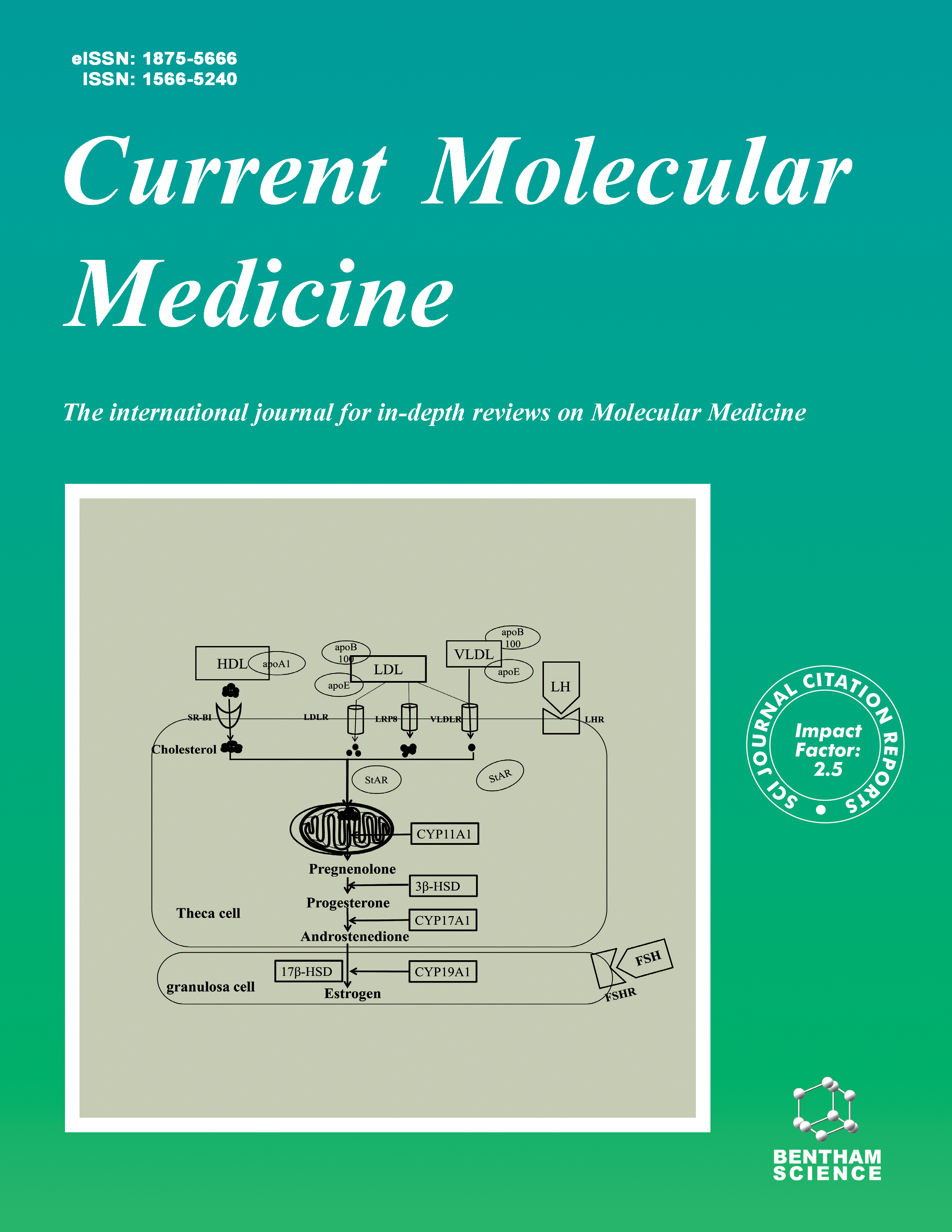
The Effects of Cholesterol Metabolism on Follicular Development and Ovarian Function
More LessAuthors: Qin Huang, Yannan Liu, Zhen Yang, Yuanjie Xie and Zhongcheng MoCholesterol is an important substrate for the synthesis of ovarian sex hormones and has an important influence on follicular development. The cholesterol in follicular fluid is mainly derived from plasma. High-density lipoprotein (HDL) and lowdensity lipoprotein (LDL) play important roles in ovarian cholesterol transport. The knockout of related receptors in the mammalian HDL and LDL pathways results in the reduction or absence of fertility, leading us to support the importance of cholesterol homeostasis in the ovary. However, little is known about ovarian cholesterol metabolism and the complex regulation of its homeostasis. Here, we reviewed the cholesterol metabolism in the ovary and speculated that regardless of the functioning of cholesterol metabolism in the system or the ovarian microenvironment, an imbalance in cholesterol homeostasis is likely to have an adverse effect on ovarian structure and function.
-
-
-
Identification of Phosphorylation Associated SNPs for Blood Pressure, Coronary Artery Disease and Stroke from Genome-wide Association Studies
More LessAuthors: Xingchen Wang, Xingbo Mo, Huan Zhang, Yonghong Zhang and Yueping ShenPurpose: Phosphorylation-related SNP (phosSNP) is a non-synonymous SNP that might influence protein phosphorylation status. The aim of this study was to assess the effect of phosSNPs on blood pressure (BP), coronary artery disease (CAD) and ischemic stroke (IS). Methods: We examined the association of phosSNPs with BP, CAD and IS in shared data from genome-wide association studies (GWAS) and tested if the disease loci were enriched with phosSNPs. Furthermore, we performed quantitative trait locus analysis to find out if the identified phosSNPs have impacts on gene expression, protein and metabolite levels. Results: We found numerous phosSNPs for systolic BP (count=148), diastolic BP (count=206), CAD (count=20) and IS (count=4). The most significant phosSNPs for SBP, DBP, CAD and IS were rs1801131 in MTHFR, rs3184504 in SH2B3, rs35212307 in WDR12 and rs3184504 in SH2B3, respectively. Our analyses revealed that the associated SNPs identified by the original GWAS were significantly enriched with phosSNPs and many well-known genes predisposing to cardiovascular diseases contain significant phosSNPs. We found that BP, CAD and IS shared for phosSNPs in loci that contain functional genes involve in cardiovascular diseases, e.g., rs11556924 (ZC3HC1), rs1971819 (ICA1L), rs3184504 (SH2B3), rs3739998 (JCAD), rs903160 (SMG6). Four phosSNPs in ADAMTS7 were significantly associated with CAD, including the known functional SNP rs3825807. Moreover, the identified phosSNPs seemed to have the potential to affect transcription regulation and serum levels of numerous cardiovascular diseases-related proteins and metabolites. Conclusion: The findings suggested that phosSNPs may play important roles in BP regulation and the pathological mechanisms of CAD and IS.
-
-
-
Effects of Follicular Helper T Cells and Inflammatory Cytokines on Myasthenia Gravis
More LessAuthors: Lifang Wang, Yu Zhang, Mingqin Zhu, Jiachun Feng, Jinming Han, Jie Zhu and Hui DengBackground: Myasthenia gravis (MG) is an autoimmune disorder mediated by antibodies against the acetylcholine receptors (AChR) of the skeletal muscles. An imbalance in various T helper (Th) cells, including Th1, Th2, Th17, Th22 and follicular helper T (TFH) cells, has been found associated with immunological disturbances. Objective: In this study, we aim to investigate the role of the Th cells in peripheral blood of MG patients. Materials and Methods: A total of 33 MG patients and 34 age matched controls were enrolled in this study. Peripheral blood mononuclear cells (PBMCs) were isolated using Ficoll-Paque density gradient centrifugation assay. The proportion of TFH cells in PBMC were analyzed using flow-cytometry assay by determining the levels of cellular markers CD4, CXCR5, CD45RO, CD45RA and ICOS and PD-1. The levels of IFN-γ, IL-4, IL-17 and IL-21 in serum were analyzed by Cytometric Bead Array. The serum IL-22 level was analyzed by ELISA. Results: The frequency of TFH cells in PBMCs was higher than those in healthy subjects and correlated to the severity of MG patients. The levels of pro-inflammatory cytokines IFN-γ, IL-17 and IL-21 were elevated in the serum of MG patients, while there were no significant differences regarding the levels of IL-4 and IL-22 between MG patients and control subjects. Conclusion: Our findings suggest that Th cells and their cytokines balance of MG patients are involved in the clinical condition or severity of MG disease.
-
-
-
Functional Changes of Paneth Cells in the Intestinal Epithelium of Mice with Obstructive Jaundice and After Internal and External Biliary Drainage
More LessAuthors: Xiaopeng Tian, Zixuan Zhang and Wen LiObjective: To investigate the functional changes of Paneth cells in the intestinal epithelium of mice with obstructive jaundice (OJ) and after internal biliary drainage (ID) and external biliary drainage (ED). Methods: The experiment was divided into two stages. First stage: Mice were randomly assigned to two groups: (I) sham operation (SH); (II) OJ. The mice were sacrificed before the operation and on the 1st, 3rd, 5th and 7th day after the operation to collect specimens. Second stage: Mice were randomly assigned to four groups: (I) SH; (II) OJ; (III) OJ and ED; and (IV) OJ and ID. They were reoperated on day 5 for biliary drainage procedure. The specimens were collected on day 10. Results: The expressions of lysozyme and cryptdin-4 increased first and then decreased over time in group OJ, and the number of Paneth cells decreased gradually with the extension of OJ time〈p<0.05. After the secondary operation on the mice to relieve OJ, the number of Paneth cells and expressions of lysozyme and cryptdin-4 in group ID increased more significantly than those in group ED〈p<0.05〉. Conclusion: OJ could cause intestinal Paneth cells to dysfunction in mice. ID was more significant than ED in restoring the function of Paneth cells. It might be one of the mechanisms that make ID superior to ED.
-
-
-
A COL4A5 Missense Variant in a Han-Chinese Family with X-linked Alport Syndrome
More LessAuthors: Yuan Wu, Yi Guo, Jinzhong Yuan, Hongbo Xu, Yong Chen, Hao Zhang, Mingyang Yuan, Hao Deng and Lamei YuanBackground: Alport syndrome (AS) is an inherited familial nephropathy, characterized by progressive hematuric nephritis, bilateral sensorineural hypoacusis and ocular abnormalities. X-linked AS (XLAS) is the major AS form and is clinically heterogeneous, and it is associated with defects in the collagen type IV alpha 5 chain gene (COL4A5). Objective: The purpose of this research is to detect the genetic defect responsible for renal disorder in a 3-generation Han-Chinese pedigree. Methods: Detailed family history and clinical data of the family members were collected and recorded. Whole exome sequencing (WES) was applied in the proband to screen potential genetic variants, and then Sanger sequencing was used to verify the variant within the family. Two hundred unrelated ethnically matched normal individuals (male/female: 100/100, age 37.5 ± 5.5 years) without renal disorder were recruited as controls. Results: Three patients (I:1, II:1 and II:2) presented microscopic hematuria and proteinuria, and the patient I:1 developed uremia and end stage renal disease (ESRD) by age 55 and showed sensorineural hearing loss. Patient II:2 developed mild left ear hearing loss. Cataracts were present in patients I:1 and II:1. A COL4A5 gene missense variant, c.2156G>A (p.G719E), located in the Gly-X-Y repeats of exon 28, was identified to co-segregate with the renal disorder in this family. The variant was absent in 200 ethnically matched controls. Conclusion: By conducting WES and Sanger sequencing, a COL4A5 missense variant, c.2156G>A (p.G719E), was identified to co-segregate with the renal disorder, and it is possible that this variant is the genetic cause of the disorder in this family. Our study may extend the mutation spectrum of XLAS and may be useful for genetic counseling of this family. Further functional studies associated with genetic deficiency are warranted in the following research.
-
-
-
Dynamic Changes of RFRP3/GPR147 in the Precocious Puberty Model Female Rats
More LessAuthors: Wen Sun, Suhuan Li, Zhanzhuang Tian, Yumin Shi, Jian Yu, Yanyan Sun and Yonghong WangBackground: Pubertal development is a complex physiological process regulated by the neuroendocrine system and hypothalamic-pituitary-gonadal axis. Sexual precocity is a common childhood endocrine disease.The pathogenesis of sexual precocity has not been fully elucidated. RFRP3/GPRl47 signal pathway is able to inhibit the reproductive capability in avians and mammals, probably by acting on the GnRH neuron and pituitary to regulate gonadotrophin synthesis and release. However, little is known about the role of RFRP3 in puberty development and sexual precocity. Objective: To observe the dynamic changes of RFamide related peptide 3/G proteincoupled receptor 147 (RFRP3/GPR147) in hypothalamic during puberty development and explore their role in precocious puberty based on a female rat model. Methods: The Sprague-Dawley female rats were randomly divided into three groups, normal, vehicle, and precocious puberty model. At 5 days old, the rat model with precocious puberty was prepared by subcutaneously injecting a mixture of danazoldissolved ethanol and glycol. At different day-age (15, 25, 30, 35, and 40 days), the levels of estradiol(E2), follicle-stimulating hormone(FSH), and luteinizing hormone (LH) in the peripheral blood were detected by the enzyme-linked immunosorbent assay, the messenger ribonucleic acid (mRNA) expressions of RFRP3, gonadotropin releasing hormone and GPR147 were examined by real-time polymerase chain reaction(R-T PCR). RFRP3 positive cells were observed using Immunofluorescence confocal microscopy. Results: At 25 and 30 days, the levels of sex hormones and the uterus coefficients were significantly higher in the precocious puberty model group than those in the normal and vehicle groups. The ovarian morphological development in the precocious puberty model rats was significantly earlier than those in the normal and vehicle groups. The mRNA expressions of RFRP3/GPR147 and GnRH in the precocious puberty model group gradually increased and peaked at 25 days. The different day-age and the interaction have significant statistical significance on the expression of RFRP3 mRNA, while the levels of RFRP3 mRNA in the model group and vehicle groups have no significant statistical significance. There was statistical significance between the model group and vehicle groups in different day-age on the expression of GPR147 mRNA.The expression of hypothalamic RFRP3/GPR147 mRNA and RFRP3 positive cells gradually decreased with puberty onset. At 35 days, the levels of RFRP3 mRNA and GPR147 mRNA were significantly lower in the precocious puberty model group than those in the vehicle groups. Meanwhile, the levels of LH in the precocious puberty model rats reached its peak at this age. In the vehicle group, the levels of RFRP3 mRNA and serum LH were gradually increased and LH nearly peaked at 35 day-age. Subsequently, it gradually decreased and reached the lowest level at 35 day-age. The expression of RFRP3 mRNA and LH were positively correlated. Conclusion: The findings suggested that RFRP3/GPR147 signaling pathway may be involved in the pathogenesis of sexual precocity by regulating puberty development and sexual maturity in rats.
-
-
-
Frequency of Interleukins IL1ß/IL18 and Inflammasome NLRP1/NLRP3 Polymorphisms in Sickle Cell Anemia Patients and their Association with Severity Score
More LessBackground: Interleukins IL1ß/IL18 and Inflammasome NLRP1/NLRP3 polymorphisms can change the course of multiple human diseases, both inflammatory as infectious. SNPs these proteins were associated with the constructive activation of the Inflammasome and excessive production of IL-1β induce a serious autoinflammatory disease, as sickle cell anemia (SCA). The present study aims to association of interleukins IL1ß/IL18 and inflammasome NLRP1/NLRP3 polymorphisms in SCA patients in Amazon region and their association with severity score. Methods: The study was developed at Fundação Hospitalar de Hematologia e Hemoterapia do Amazonas (HEMOAM) with 21 patients diagnosed SCA (HbSS) and 50 Healthy Donor´s. Genetic polymorphisms (SNPs) in interleukins IL1ß/IL18 and inflammasome NLRP1/NLRP3 were genotyped by polymerase chain reaction-restriction fragment length polymorphism (PCR-RFLP) and real time PCR. Simple and multiple logistic regression were performed to investigate association between the polymorphisms and the SCA and severe score. Results: The genotypes C/C (IL18 -137G/C) and C/A (NLRP3, rs35829419) appear to be risk factors for SCA disease (IL18: G/G vs C/C OR=103.500 [95% CI: 8.32-1287.79, p<0.00001]; IL18: G/G vs G/C OR=7.360 [95% CI: 0.85-63.48, p=0.040]; IL18: G/G vs CC+CG OR=14.481 [95% CI: 1.79-117.32, p=0.002; NLRP3: C/C vs C/A: OR=10.967 [95% CI: 2.41-49.89, p=0.0004]). In addition, only allelic C (IL18 -137G/C) and A (NLRP3) appear to be risk factors for SCA disease (IL18: G vs C OR=6.366 [95% CI: 2.73-14.86, p<0.00001]; NLRP3: C vs A OR=8.383 [95% CI: 2.03-34.62, p=0.005]. No associations were observed between genotypes and alleles with the severity score. Conclusion: Evidence of association between the IL18 (rs16944) and NLRP3 (rs35829419) polymorphisms with sickle cell anemia were described. Our results suggest that individuals with genotypes evaluated are associated SCA disease even though it does not influence the severe score.
-
Volumes & issues
-
Volume 25 (2025)
-
Volume 24 (2024)
-
Volume 23 (2023)
-
Volume 22 (2022)
-
Volume 21 (2021)
-
Volume 20 (2020)
-
Volume 19 (2019)
-
Volume 18 (2018)
-
Volume 17 (2017)
-
Volume 16 (2016)
-
Volume 15 (2015)
-
Volume 14 (2014)
-
Volume 13 (2013)
-
Volume 12 (2012)
-
Volume 11 (2011)
-
Volume 10 (2010)
-
Volume 9 (2009)
-
Volume 8 (2008)
-
Volume 7 (2007)
-
Volume 6 (2006)
-
Volume 5 (2005)
-
Volume 4 (2004)
-
Volume 3 (2003)
-
Volume 2 (2002)
-
Volume 1 (2001)
Most Read This Month


