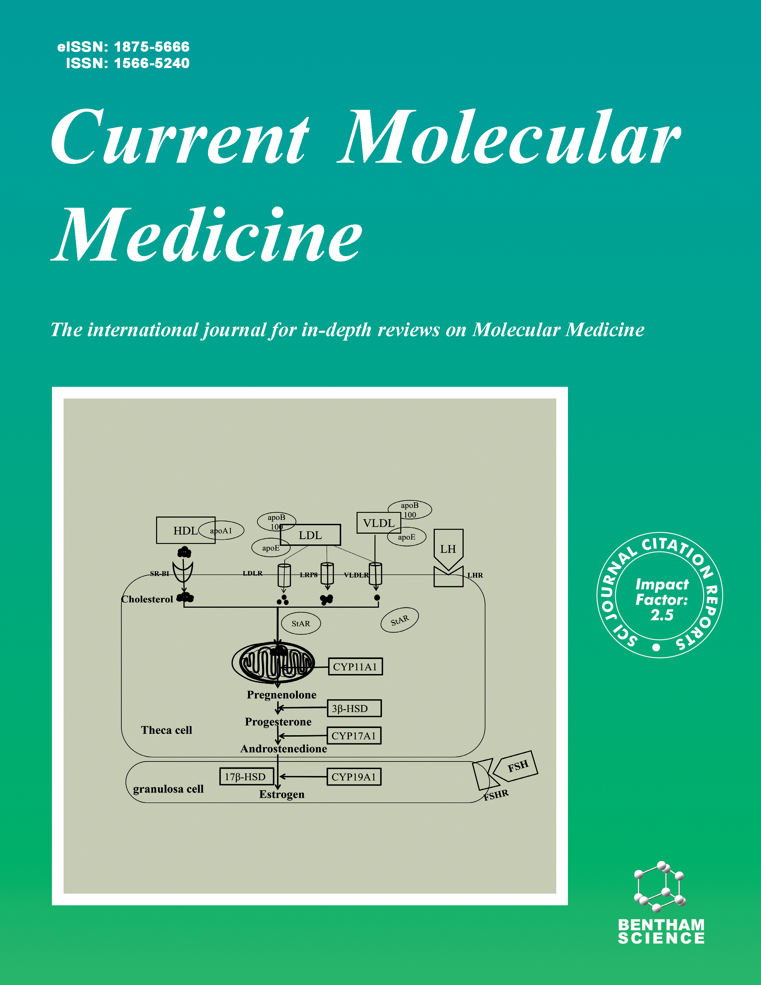
Current Molecular Medicine - Volume 18, Issue 10, 2018
Volume 18, Issue 10, 2018
-
-
Human Disease Modelling Techniques: Current Progress
More LessAuthors: Victor Yu. Glanz, Alexander N. Orekhov and Alexey V. DeykinAdvances in genetic engineering and genomic studies facilitated the development of animal models of human diseases. To date, numerous models based on different animal species are available for the most socially significant human diseases, such as cardiovascular disorders, cancer, neurodegenerative and metabolic disorders. Modern genetic methods allow creating animals with certain genes up- or downregulated, as well as bearing specific mutations. However, this precision is not easy to translate into clinical practice: animal models still have their limitations, including both physiological and genetic differences between humans and animals and complexity of disease conditions that are difficult to reproduce. In this review, we will discuss the most relevant modern techniques that allow creating genetically engineered animal models.
-
-
-
GDF11 Attenuated ANG II-Induced Hypertrophic Cardiomyopathy and Expression of ANP, BNP and Beta-MHC Through Down-Regulating CCL11 in Mice
More LessAuthors: Chenjun Zhang, Ying Wang, Zhiru Ge, Jie Lin, Jiwen Liu, Xiaofei Yuan and Zijun LinBackground: Growth differentiation factor 11 (GDF11) decreases with age, and increased C-C motif chemokine 11 (CCL11) is involved in aging. However, the effects of GDF11 on Angiotensin II (ANG II)-induced hypertrophic cardiomyopathy and expression of markers for volume overload and hypertrophy such as ANP, BNP and beta-MHC, as well as the relationship between GDF11 and CCL11 in hypertrophic cardiomyopathy are unclear. Therefore, the current study aimed to examine the effects of GDF11 on ANG II-induced hypertrophic cardiomyopathy and expression of ANP, BNP and beta-MHC in mice, and explore possible molecular mechanisms. Methods: Vectors were constructed and viruses were packaged. Mouse cardiomyocytes were treated with ANG II for 24 h. Meanwhile, mouse cardiomyocytes were divided into 4 groups: (1) control; (2) ANG II; (3) ANG II+GDF11; and (4) ANG II+CCL11. Furthermore, mouse cardiomyocytes were treated with GDF11 and CCL11 proteins for 48 h, respectively. The thickness of IVS and LVPS during systole and diastole were measured by cardiac ultrasound in the mouse model of hypertrophic cardiomyopathy. The relative expression of ANP, BNP, beta-MHC, CCL11 and GDF11 in cardiomyocytes or heart tissue of mice was detected by qPCR or Western blot. 3’- UTR luciferase reporter assay was utilized to examine the relationship between GDF11 and the expression of CCL11. Results: The expression of ANP, BNP, and beta-MHC in mouse cardiomyocytes was significantly increased after the cells were treated with 800 nM ANG II, which was utilized in the following cell experiments. After ANG II treatment, 0.2 ng/ml GDF11 group displayed the highest inhibition of expression of ANP, BNP and beta-MHC in mouse cardiomyocytes, whereas 50 ng/ml CCL11 group displayed the highest stimulation of the expression. GDF11 at 10 ng/ml significantly decreased the expression of CCL11 in mouse cardiomyocytes as compared to the control group. Mice treated with ANG II had increased thickness of IVS and LVPS during both systole and diastole, which was significantly attenuated by GDF11 overexpression. GDF11 overexpression attenuated the increase in expression of ANP, BNP and beta-MHC in the mice model of hypertrophic cardiomyopathy. The relative serum concentration of GDF11 was markedly decreased, and CCL11 was dramatically increased in mice with hypertrophic cardiomyopathy. GDF11 overexpression restored the serum concentration of GDF11 and CCL11 in the mice model of hypertrophic cardiomyopathy. In addition, GDF11 interference group had markedly increased expression of CCL11, whereas GDF11 overexpression group had significantly decreased expression of CCL11 in luciferase reporter assay. Conclusions: GDF11 attenuated ANG II-induced hypertrophic cardiomyopathy and expression of ANP, BNP and beta-MHC through down-regulating CCL11 in mice.
-
-
-
Estriol Inhibits Dermcidin Isoform-2 Induced Inflammatory Cytokine Expression Via Nitric Oxide Synthesis in Human Neutrophil
More LessAuthors: Pradipta Jana, Md. M. Khan, Subrata Kumar De, Asru K. Sinha, Santanu Guha, Gausal A. Khan and Smarajit MaitiBackground: An increase in the level of cytokines like TNF-α and IL-6 causes the inflammatory surge in acute ischemic heart disease (AIHD). Objective: A high-level dermcidin isoform-2 (DCN-2) occurrence in AIHD was subjected to determine a possible regulation of cytokines expression. The effect of estrogen to counteract the inflammatory response was determined. Methods: Blood was collected from AIHD patients and normal volunteers with consent. Nitric oxide (NO) synthesis was done with methemoglobin method.TNF-α and IL-6 expression were determined by ELISA and Western blot. Results: (DCN-2) incubation with 120nM to the normal neutrophil solution for 2h resulted in the increase of TNF-α from 3.82±1.53pg/ml to 20.7±6.9pg/ml and IL-6 from 3.27±1.52pg/ml to 47.07±3.4pg/ml. In AIHD patients, the cytokine level was18.3- 27.3pg/ml, with a median value 21.86pg/ml (TNF-α) and IL-6 level was 23.54- 52.73pg/ml, with a median value 42.16pg/ml. Treatment with 0.6nM estriol, a kind of female steroid hormone estrogen for 45min decreased the elevated cytokine level in 120nM DCN-2 treated normal neutrophils. DCN-2 induced TNF-α synthesis in neutrophils was further determined by Western blot technique with a thickened band intensity of TNF-α. Estriol (0.6nM) treatment also influenced the DCN-2 induced inhibition of nitric oxide (NO) synthesis from 0nmol NO/ml to 0.56nmol/ml. The subsequent reduction of TNF-α level correlates the increase of NO level. Conclusion: In conclusion, the stress-induced DCN-2 production in AIHD propagates the inflammatory response. Steroid molecule like estriol plays a protective role by reducing DCN-2 responses in the NO synthesis.
-
-
-
Th1/Th17 Cytokine Profile is Induced by Macrophage Migration Inhibitory Factor in Peripheral Blood Mononuclear Cells from Rheumatoid Arthritis Patients
More LessAuthors: Samuel García-Arellano, Luis A. Hernández-Palma, Richard Bucala, Jorge Hernández-Bello, Ulises De la Cruz-Mosso, Trinidad García-Iglesias, Sergio Cerpa-Cruz, Jorge Ramón Padilla-Gutiérrez, Yeminia Valle, José Guadalupe Soñanez-Organis, Isela Parra-Rojas, Ana Laura Pereira-Suárez and José Francisco Muñoz-ValleBackground: Macrophage migration inhibitory factor (MIF) is an immunoregulatory cytokine that plays a crucial role as a regulator of the innate and adaptive immune responses and takes part in the destructive process of the joint in rheumatoid arthritis (RA) by promoting angiogenesis and inducing proinflammatory cytokines and matrix metalloproteinases (MMP). We evaluated if recombinant human MIF (rhMIF) induces the production of TNF-α, IFN-γ, IL-1β, IL-6, IL-10, IL-17A, and IL- 17F in peripheral blood mononuclear cells (PBMC) from RA patients and control subjects (CS). Methods: The PBMC from RA patients and CS were stimulated for 24 hours with combinations of LPS, rhMIF or the MIF antagonist ISO-1. Cytokine profiles were measured using a multiplex immunoassay and, macrophage migration inhibitory factor (MIF) was determined by ELISA kit. Results: The PBMC of CS and RA produced Th1 and Th17 cytokines under stimulation with rhMIF, however, this effect was higher in the cells of RA patients. The rhMIFstimulated PBMC from RA patients produced higher levels of Th1 and Th17 cytokines in comparison with unstimulated cells: TNF-α (538.81 vs. 5.02 pg/mL, p<0.001), IFN-γ (721.90 vs. 8.40 pg/mL, p<0.001), IL-1β (150.14 vs. 5.17 pg/mL, p<0.05), IL-6 (19769.70 vs. 119.85 pg/mL, p<0.001), IL-17A (34.97 vs. 0.90 pg/mL, p<0.01) and IL-17F (158.43 vs. 0.92 pg/mL, p<0.001). Conclusion: These results highlight the potential role of MIF in the establishment of the chronic inflammatory process in RA via Th1 and Th17 cytokine profile induction and provide new evidence of the role of MIF to stimulate the IL-17A and IL-17F expression in PBMC from RA and CS.
-
-
-
Potential Mutations in Chinese Pathologic Myopic Patients and Contributions to Phenotype
More LessPurpose: Pathologic myopia is a leading cause of visual impairment in East Asia. The aim of this study was to investigate the potential mutations in Chinese pathologic myopic patients and to analyze the correlations between genotype and clinical phenotype. Methods: One hundred and three patients with pathologic myopia and one hundred and nine unrelated healthy controls were recruited from Zhongshan Ophthalmic Center. Detailed clinical data, including ultra-widefield retinal images, measurements of bestcorrected visual acuity, axial length, refractive error and ophthalmic examination results, were obtained. Blood samples were collected for high-throughput DNA targeted sequencing. Based on the screening results, phenotype-genotype correlations were analyzed. Results: The study included 196 eyes of 103 patients (36 men and 67 women) with an average age of 52.19 (38.92 – 65.46) years, an average refractive error of -11.80 D (- 16.38 – -7.22) and a mean axial length of 28.26 mm (25.79 – 30.73). The patients were subdivided into three groups: myopic chorioretinal atrophy (190 eyes of 101 patients), myopic choroidal neovascularization (17 eyes of 15 patients), and myopic traction retinopathy (71 eyes of 61 patients). Systematic analysis of variants in the 255 genes revealed six potential pathogenic mutations: PEX7, OCA2, LRP5 (rs545382, c.1647T>C), TSPAN12 (rs41623, c.765G>T), RDH5 (rs3138142, c.423C>T) and TTC21B (rs80225158, c.2385G>C). OCA2 mutations were primarily observed in patients with myopic traction maculopathy. Conclusion: Genetic alterations contribute to various clinical characteristics in Chinese pathologic myopic patients. The study may provide new insights into the etiology of pathologic myopia and potential targets for therapeutic interventions.
-
-
-
Naoxintong Retards Atherosclerosis by Inhibiting Foam Cell Formation Through Activating Pparα Pathway
More LessAuthors: Zeng Wang, Huairui Shi, Huan Zhao, Zhen Dong, Buchang Zhao, Xinyu Weng, Rongle Liu, Xiao Li, Kai Hu, Yunzeng Zou, Aijun Sun and Junbo GeBackgrounds: We recently reported that Naoxintong (NXT), a China Food and Drug Administration (FDA)-approved cardiac medicine, could reduce the plaque size, but the underlying mechanism remains elusive now. Objective: In this study, we investigated the effects of NXT on foam cell accumulation both in vivo and in vitro and explored related mechanisms. Method: THP-1 cells and bone marrow-derived macrophages were incubated with oxidized low-density lipoprotein (ox-LDL) with/without Naoxintong. ApoE-/- mice fed an atherogenic diet were administered to receive NXT for eight weeks. Macrophage-derived foam cell formation in plaques was measured by immunohistochemical staining. Expression of proteins was evaluated by Western blot. Lentivirus was used to knockdown PPARα in THP-1 cells. Results: After NXT treatment, foam cell accumulation was significantly reduced in atherosclerotic plaques. Further investigation revealed that oxidized low-density lipoprotein (ox-LDL) uptake was significantly decreased and expression of scavenger receptor class A (SR-A) and class B (SR-B and CD36) was significantly downregulated post-NXT treatment. On the other hand, NXT increased cholesterol efflux and upregulated ATP-binding cassette (ABC) transporters (ABCA-1 and ABCG-1) in macrophages. Above beneficial effects of NXT were partly abolished after lentiviral knockdown of PPARα. Conclusion: Our findings suggest that NXT could retard atherosclerosis by inhibiting foam cell formation through reducing ox-LDL uptake and enhancing cholesterol efflux and above beneficial effects are partly mediated through PPARα pathway.
-
-
-
Histone Deacetylase Inhibition Attenuates Cardiomyocyte Hypoxia-Reoxygenation Injury
More LessBackground: Cardiac reperfusion injury can have devastating consequences. Histone deacetylase (HDAC) inhibitors are potent cytoprotective agents, but their role in the prevention of cardiac injury remains ill-defined. Objective: We sought to determine the therapeutic potential of HDAC inhibitors in an in vitro model of cardiomyocyte hypoxia-reoxygenation (H/R). Method: H9c2 cardiomyocytes were subjected to H/R and treated with various classspecific and pan-HDAC inhibitors in equal concentrations (5μM). Biological activity of inhibitors was determined, as a proxy for concentration adequacy, by Western blot for acetylated histone H3 and α-tubulin. Cell viability and cytotoxicity were measured by methyl thiazolyl tetrazolium and lactate dehydrogenase assays, respectively. Mechanistic studies were performed to better define the effects of the most effective agent, Tubastatin-A (Tub-A), on the phosphoinositide 3-kinase (PI3K)/mammalian target of rapamycin (mTOR) pathway effectors, and on the degree of autophagy. Results: All inhibitors acetylated well-known target proteins (histone H3 and α-tubulin), suggesting that concentrations were adequate to induce a biological effect. Improved cell viability and decreased cell cytotoxicity were noted in cardiomyocytes exposed to Tub-A, whereas the cytoprotective effects of other HDAC inhibitors were inconsistent. Pro-survival mediators in the PI3K/mTOR pathway were up-regulated and the degree of autophagy was significantly attenuated in cells that were treated with Tub-A. Conclusion: HDAC inhibitors improve cell viability in a model of cardiomyocyte H/R, with Class IIb inhibition (Tub-A) demonstrating superior cellular-level potency and effectiveness. This effect is, at least in part, related to an increased expression of prosurvival mediators and a decreased degree of autophagy.
-
Volumes & issues
-
Volume 25 (2025)
-
Volume 24 (2024)
-
Volume 23 (2023)
-
Volume 22 (2022)
-
Volume 21 (2021)
-
Volume 20 (2020)
-
Volume 19 (2019)
-
Volume 18 (2018)
-
Volume 17 (2017)
-
Volume 16 (2016)
-
Volume 15 (2015)
-
Volume 14 (2014)
-
Volume 13 (2013)
-
Volume 12 (2012)
-
Volume 11 (2011)
-
Volume 10 (2010)
-
Volume 9 (2009)
-
Volume 8 (2008)
-
Volume 7 (2007)
-
Volume 6 (2006)
-
Volume 5 (2005)
-
Volume 4 (2004)
-
Volume 3 (2003)
-
Volume 2 (2002)
-
Volume 1 (2001)
Most Read This Month


