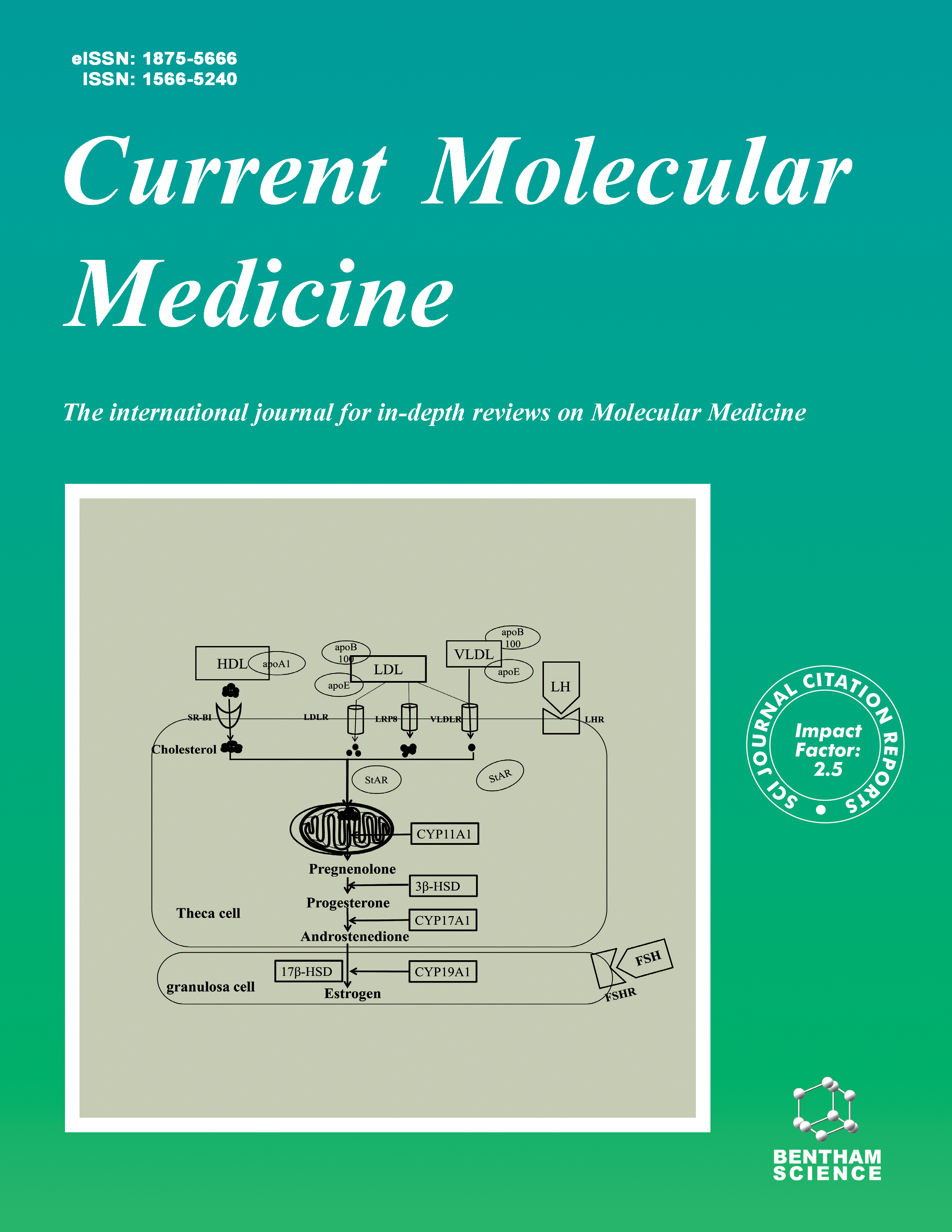
Current Molecular Medicine - Volume 17, Issue 7, 2017
Volume 17, Issue 7, 2017
-
-
Elevated Interleukin 37 Expression Associated With Disease Activity in HLA-B27 Associated Anterior Uveitis and Idiopathic Anterior Uveitis
More LessBackground & Objective: Interleukin 37 (IL-37) is an important regulator of the anti-inflammatory T-cell response. In this study, we investigated its expression and function in peripheral blood mononuclear cells (PBMCs) of patients with HLA-B27 associated acute anterior uveitis (AAU) and idiopathic AAU. Methods: 15 patients with HLA-B27-associated AAU, 10 patients with idiopathic AAU and 22 controls were recruited to this study from August 2013 to December 2016. Complete ophthalmological examinations were performed and clinical features were clearly documented. Blood samples were collected and peripheral blood mononuclear cells were extracted. IL-37 messenger RNA (mRNA) and protein expression in peripheral blood mononuclear cells (PBMCs) were examined by performing RT-PCRs and western blot, respectively. Cytokines in the supernatants of stimulated dendritic cells (DCs) with IL-37 were assayed by multiplex immunoassay. Results: An increased level of IL-37 mRNA and protein expression by PBMCs was found in the patient group with clinically active AAU compared to controls. There was no significant difference in IL-37 mRNA and protein expression levels between HLA-B27 associated AAU and idiopathic AAU. IL-37 significantly inhibited the production of IL-1β, IL-6, IL-10, IL-21, IL-23, TNF-α and IFN-γ. IL-37 levels of mRNA and protein expression showed a significant positive correlation with disease activity. Conclusions: Elevated IL-37 expression is associated with disease activity in HLA-B27 associated AAU and idiopathic AAU. IL-37 can inhibit proinflammatory cytokine productions in AAU. Manipulation of IL-37 may offer a new therapeutic target for these entities.
-
-
-
How To Deal With Uveitis Patients?
More LessDuring the past nine years, our center has grown into the largest uveitis referral center in China. To deal with this increasing stream of patients we have developed a management system to coordinate communication with our patients, their referring ophthalmologists, consultations with other medical specialties and worldrenowned foreign uveitis specialists. We have established the biggest database of uveitis patients records allowing continuous analysis of clinical features and response to treatment of patients with various uveitis entities as well as the evaluation of the relevance of various ancillary tests performed in this patient group. The establishment of a specimen biobank has been shown to be instrumental in the research on the complex immunopathological mechanisms involved in this disease. The close interaction between patient care and clinical research under one roof has led to a novel understanding of disease mechanisms and will undoubtedly lead to a tailored treatment for this disease.
-
-
-
Association of IL33 and IL1RAP Polymorphisms With Acute Anterior Uveitis
More LessAuthors: X.-F. Huang, W. Chi, D. Lin, M.-L. Dai, Y.-L. Wang, Y.-M. Yang, Z.-B. Jin and Y. WangBackground: AAU (acute anterior uveitis) is the most common entity of uveitis characterized by acute vision loss and violent sore eyes. IL-33 and IL-1RacP have been found to play crucial roles in the innate immune system. Objective: In the present study, we investigated the association of IL33 and IL1RAP genes with AAU. Method: A total of 549 AAU patients and 1080 unrelated healthy controls were recruited for this study. Ten single nucleotide polymorphisms (SNPs) were genotyped using Sequenom Mass ARRAY technology. Results: Our findings demonstrated that IL1RAP-rs3773978 significantly associated with AAU and could serve as a genetic risk marker in Chinese AAU patients. A significantly increased frequency of the A allele and AA homozygosity of IL1RAP-rs3773978 was observed in AAU patients compared with that in controls (p=0.001, pc=0.01, OR=1.282, 95% CI 1.106 to 1.487; p=0.0003, pc=0.003, OR=1.647, 95% CI 1.255 to 2.163, respectively). Further stratification analyses showed that the genetic correlation may differ depending on HLA-B27 status, AS (ankylosing spondylitis) status, attack times and laterality status. Conclusion: Our findings provide new insights that enhance the current knowledge of uveitis genetics by demonstrating the specific functional roles of IL1RAP and other IL-1 family genes in uveitis.
-
-
-
Mesenchymal Stem Cells Inhibited Dendritic Cells Via the Regulation of STAT1 and STAT6 Phosphorylation in Experimental Autoimmune Uveitis
More LessPurpose: We have previously reported that MSCs inhibited experimental autoimmune uveitis (EAU) in rodent models induced by either uveitogenic antigens or antigen-specific T cells. In this study, we explored the inhibitory mechanisms of MSCs on dendritic cells (DCs) in EAU. Methods: We collected the DCs from the lymph nodes of MSC treated or untreated EAU rats, as well as bone marrow derived DCs cultured in vitro with or without MSC treatment. The levels of costimulatory molecules of CD80, CD86, CD40, OX40L and suppressors of cytokine signaling (SOCS1, SOCS2, and SOCS3) on these DCs were analyzed by flow Cytometry. The expression of CCR-7 and MMP-9 was examined by real time PCR and western blots. Total proteins of STAT1 and STAT6 signaling molecules and their phosphorylation were examined by western blots. ShRNA of STAT1 and STAT6 were respectively employed to explore the influence of STAT1 and STAT6 knockdown on DCs. Results: MSC treatment down-regulated the expression of CD80, CD86, CD40, and OX40L, as well as CCR-7 and MMP-9, but increased the levels of SOCS1, SOCS 2, and SOCS3 on DCs. STAT1 phosphorylation was reduced while STAT6 phosphorylation was enhanced in MSC treated DCs. Moreover, MSC treatment and STAT1 shRNA equally reduced CCR-7 and MMP-9 levels in DCs, and inhibited the proliferation of R16-specific T cells. In contrast, knockdown of STAT6 in DCs by STAT6 shRNA increased the expression of CD80 and CD86 and accelerated the proliferation of R16-specific T cells. Conclusion: MSCs inhibit DC maturation by regulating Stat1 and Stat6 phosphorylation in EAU.
-
-
-
Puerarin Stimulates Osteogenic Differentiation and Bone Formation Through the ERK1/2 and p38-MAPK Signaling Pathways
More LessBackground: Osteoporosis is a world-wide health problem, which leads to decreased bone strength and increased susceptibility to fractures. Puerarin, a phytoestrogen extracted from Pueraria lobata (Willd.) Ohwi, has been identified as a promising intervention for preventing bone loss and promoting bone regeneration. However, the underlying mechanisms for its anabolic action are still not clear. In the present study, we aimed to investigate the effect of puerarin on the osteogenic differentiation of bone marrow stromal cells (BMSCs) and the possible molecular mechanism mediating its action. Methods: Bone marrow stromal cells (BMSCs) and intragastric administration on ovariectomized(OVX) rats were used to study the anti-osteoporotic function of puerarin. The involvement of mitogen-activated protein kinase (MAPK) signaling pathways was determined. Results: Our results demonstrated that at optimal concentration, puerarin could promote osteogenic differentiation of BMSCs in vitro. This induction was mediated by MAPK signaling pathway. Further detailed study revealed that ERK1/2-Runx2 signaling pathway had more prominent effect than p38 signaling pathway in puerarin-induced differentiation of BMSCs toward the osteogenic phenotype. We also found that puerarin protected against reduction in bone mineral density and improved femur trabecular bone structure in ovariectomized rats. Conclusion: Our findings revealed the functional mechanism of puerarin in promoting osteogenic differentiation which involved ERK1/2 and p38-MAPK pathway and provided experimental evidence for the potential application of puerarin for estrogen replacement therapy of osteoporosis.
-
-
-
Neuronal Expression of Junctional Adhesion Molecule-C is Essential for Retinal Thickness and Photoreceptor Survival
More LessBackground: Photoreceptor cell death is a key pathology of retinal degeneration diseases. To date, the molecular mechanisms for this pathological process remain largely unclear. Junctional adhesion molecule-c (Jam-c) has been shown to play important roles in different biological events. However, its effect on retinal neuronal cells is unknown. Objective: To determine the effect of Jam-c on adult mouse eyes, particularly, on retinal structure, vasculature and photoreceptor cells, in order to explore potential important target molecules for ocular diseases. Methods: Jam-c global knockout mice, endothelial-specific and neuronal-specific Jam-c conditional knockout mice using Tie2-Cre and Nestin-Cre mice respectively were used in this study. Mouse eyes were harvested from the different groups and eye size examined. Cryosections of the eyes were made and stained with Hematoxylin and Eosin (H&E) and the thicknesses of retinal layers measured. Retinal blood vessels and cone and rod photoreceptors were analyzed using isolectin B4, peanut agglutinin and rhodopsin as markers respectively. In vivo Jam-c knockdown in mouse eyes was performed by intravitreal injection of Jam-c shRNA. Jam-c expression in the retinae was quantified by real-time PCR. Results: Global Jam-c gene deletion in mice resulted in smaller eyes and decreased the diameters of lens and iris. Jam-c-/- mice display marked thinning of the outer nuclear layer (ONL), less numbers of photoreceptor cells, and abnormal retinal vasculature. Importantly, neuronal-specific Jam-c deletion led to similar phenotype, whereas no obvious defect was observed in endothelial-specific Jam-c knockout mice. Moreover, Jam-c knockdown by shRNA also decreased ONL thickness and photoreceptor numbers. Conclusion: We found that Jam-c is critically required for the normal size and retinal structure. Particularly, Jam-c plays important roles in maintaining the normal retinal thickness, vasculature and photoreceptor numbers. Jam-c thus may therefore have important roles in various ocular diseases.
-
-
-
The bHLH Protein Nulp1 is Essential for Femur Development Via Acting as a Cofactor in Wnt Signaling in Drosophila
More LessBackground: The basic helix-loop-helix (bHLH) protein families are a large class of transcription factors, which are associated with cell proliferation, tissue differentiation, and other important development processes. We reported that the Nuclear localized protein-1 (Nulp1) might act as a novel bHLH transcriptional factor to mediate cellular functions. However, its role in development in vivo remains unknown. Methods: Nulp1 (dNulp1) mutants are generated by CRISPR/Cas9 targeting the Domain of Unknown Function (DUF654) in its C terminal. Expression of Wg target genes are analyzed by qRT-PCR. We use the Top-Flash luciferase reporter assay to response to Wg signaling. Results: Here we show that Drosophila Nulp1 (dNulp1) mutants, generated by CRISPR/Cas9 targeting the Domain of Unknown Function (DUF654) in its C terminal, are partially homozygous lethal and the rare escapers have bent femurs, which are similar to the major manifestation of congenital bent-bone dysplasia in human Stuve- Weidemann syndrome. The fly phenotype can be rescued by dNulp1 over-expression, indicating that dNulp1 is essential for fly femur development and survival. Moreover, dNulp1 overexpression suppresses the notch wing phenotype caused by the overexpression of sgg/GSK3β, an inhibitor of the canonical Wnt cascade. Furthermore, qRT-PCR analyses show that seven target genes positively regulated by Wg signaling pathway are down-regulated in response to dNulp1 knockout, while two negatively regulated Wg targets are up-regulated in dNulp1 mutants. Finally, dNulp1 overexpression significantly activates the Top-Flash Wnt signaling reporter. Conclusion: We conclude that bHLH protein dNulp1 is essential for femur development and survival in Drosophila by acting as a positive cofactor in Wnt/Wingless signaling.
-
-
-
Identification of a Good-Prognosis IDH-Mutant-Like Population of Patients with Diffuse Gliomas
More LessBackground: Isocitrate dehydrogenase (IDH) mutation is the initiating event that defines major clinical and prognostic classes of gliomas, but the potential mechanisms have not been well interpreted yet. The main objective of the current study was to better understand the underlying biology of IDH mutant gliomas as captured by gene expression profiles. Methods: RNA sequencing data of WHO grade II-IV gliomas from the Chinese Glioma Genome Atlas (CGGA, N=325) were used to assess differentially expressed genes between IDH mutant and wild type gliomas and to construct a gene expression-based classifier to detect IDH mutant samples with high sensitivity and specificity. The classifier was validated in independent RNA sequencing data from the Cancer Genome Atlas (TCGA, N=699), and the prognostic value of the classifier was also assessed in the two datasets. Results: A 58-gene-pair IDH mutation signature was developed by using the top scoring pairs algorithm. In CGGA dataset, 98.5% and 100% IDH mutant samples were also predicted to be mutant by gene expression based IDH status in grade II-III and grade IV gliomas, respectively. In TCGA dataset, the proportions were 99.8% and 100%, respectively. The signature remained to be a prognostic marker in multivariate cox analysis both in CGGA and TCGA datasets. Conclusion: A characteristic gene expression signature is associated with and accurately predicts IDH mutation status. This suggests a common biology between these tumors and adds prognostic and biologic information that is not captured by the mutation status alone. These results may help in population stratification for clinical trials. As RNA-seq is more and more prevalent and cost-effective in glioma molecular diagnosis, this gene signature would provide a precise method to predict IDH mutation status with RNA-seq data.
-
Volumes & issues
-
Volume 25 (2025)
-
Volume 24 (2024)
-
Volume 23 (2023)
-
Volume 22 (2022)
-
Volume 21 (2021)
-
Volume 20 (2020)
-
Volume 19 (2019)
-
Volume 18 (2018)
-
Volume 17 (2017)
-
Volume 16 (2016)
-
Volume 15 (2015)
-
Volume 14 (2014)
-
Volume 13 (2013)
-
Volume 12 (2012)
-
Volume 11 (2011)
-
Volume 10 (2010)
-
Volume 9 (2009)
-
Volume 8 (2008)
-
Volume 7 (2007)
-
Volume 6 (2006)
-
Volume 5 (2005)
-
Volume 4 (2004)
-
Volume 3 (2003)
-
Volume 2 (2002)
-
Volume 1 (2001)
Most Read This Month


