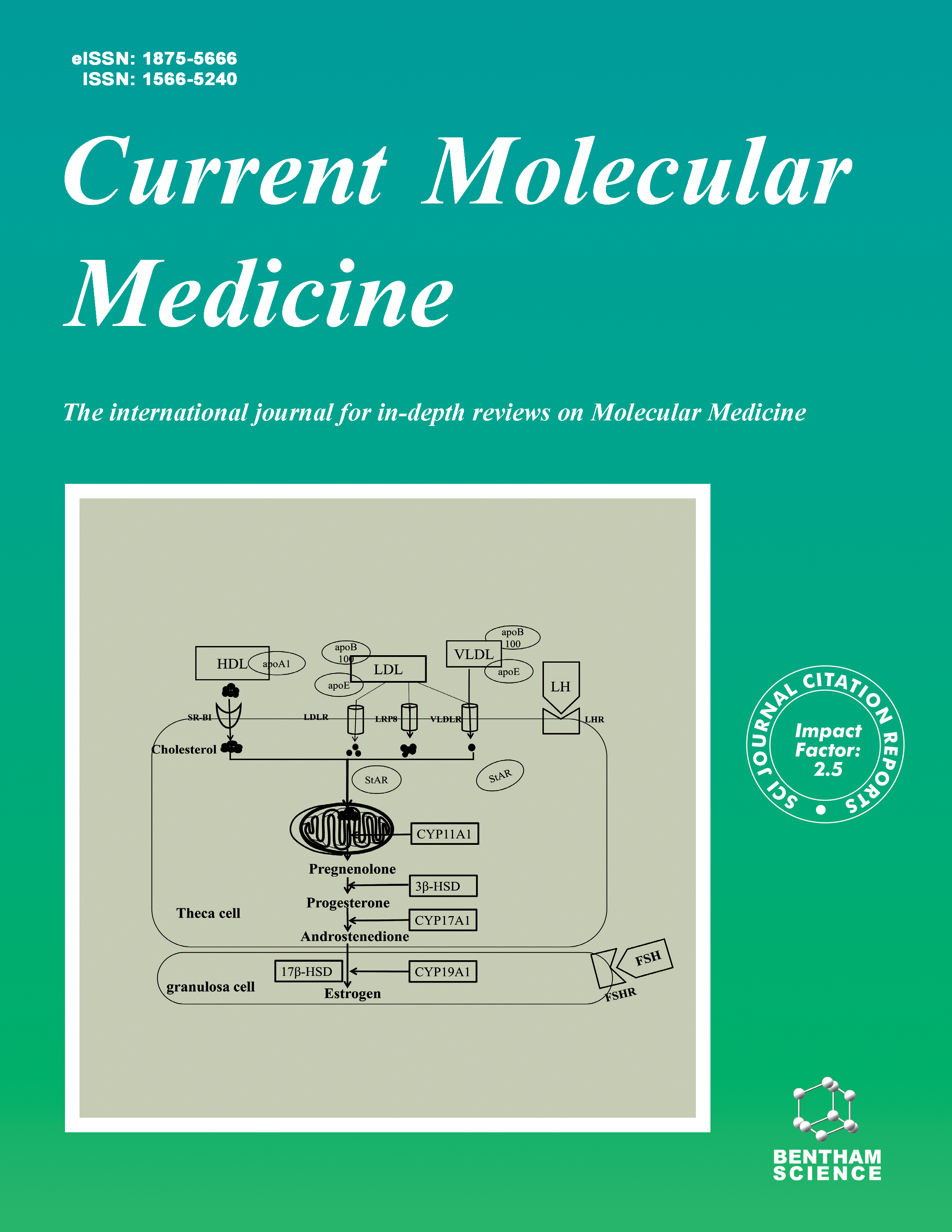
Current Molecular Medicine - Volume 16, Issue 4, 2016
Volume 16, Issue 4, 2016
-
-
HOX Genes as Potential Markers of Circulating Tumour Cells
More LessAuthors: R. Morgan and M. El-TananiCirculating tumour cells (CTCs) have significant diagnostic potential as they can reflect both the presence and recurrence of a wide range of cancers. However, this potential continues to be limited by the lack of robust and accessible isolation technologies. An alternative to isolation might be their direct detection amongst other peripheral blood cells, although this would require markers that allow them to be distinguished from an exceptionally high background signal. This review assesses the potential role of HOX genes, a family of homeodomain containing transcription factors with key roles in both embryonic development and oncogenesis, as unique and possibly disease specific markers of CTCs.
-
-
-
Prostacyclin, Atherothrombosis and Diabetes Mellitus: Physiologic and Clinical Considerations
More LessAuthors: J. Stitham and J. HwaProstacyclin (PGI2) and other metabolites of arachidonic acid are increasingly recognized for their role in the pathophysiology of human disease. A growing body of evidence from randomized controlled trials, studies of human prostacyclin receptor (hIP) variants, and IP-receptor knockout studies in mice has shown that PGI2 may have a protective effect on atherothrombotic risk. Increased risk of atherosclerosis and thrombotic sequelae may be attributed, in part, to downregulation of the prostacyclin pathway. Clinical studies with nonsteroidal antiinflammatory drugs (NSAIDs) that were selective for the cyclooxygenase-2 (COX-2) isoenzyme, although protective of mucosa in the gastrointestinal tract, first alluded to a potential role of PGI2 in atherothrombotic risk. Outcomes from early clinical trials showed a 2- to 3-fold increase in risk of incurring a thrombotic event (e.g., myocardial infarction or stroke). Further analyses suggested that atherothrombotic risk is a continuous variable with relative NSAID COX-2 selectivity, and that the COX-2 metabolic product, PGI2, appears to play a key role. Effects of reduced PGI2 levels may be felt in particular by patients with diabetes mellitus, a patient population at the high end of the cardiovascular risk spectrum. Therapies that spare PGI2 may provide the greatest level of protection. The mechanism of protection by PGI2 is under intense investigation.
-
-
-
The Impact of CRISPR/Cas9-Based Genomic Engineering on Biomedical Research and Medicine
More LessAuthors: D.E. Go and R.W. StottmannThere has been prolonged and significant interest in manipulating the genome for a wide range of applications in biomedical research and medicine. An existing challenge in realizing this potential has been the inability to precisely edit specific DNA sequences. Past efforts to generate targeted double stranded DNA cleavage have fused DNA-targeting elements such as zinc fingers and DNA-binding proteins to endonucleases. However, these approaches are limited by both design complexity and inefficient, costineffective operation. The discovery of CRISPR/Cas9, a branch of the bacterial adaptive immune system, as a potential genomic editing tool holds the promise of facile targeted cleavage. Its novelty lies in its RNA-guided endonuclease activity, which enhances its efficiency, scalability, and ease of use. The only necessary components are a Cas9 endonuclease protein and an RNA molecule tailored to the gene of interest. This lowbarrier of adoption has facilitated a plethora of advances in just the past three years since its discovery. In this review, we will discuss the impact of CRISPR/Cas9 on biomedical research and its potential implications in medicine.
-
-
-
The High Mobility Group A1 (HMGA1) Transcriptome in Cancer and Development
More LessAuthors: T.F. Sumter, L. Xian, T. Huso, M. Koo, Y.-T. Chang, T.N. Almasri, L. Chia, C. Inglis, D. Reid and L.M.S. ResarBackground & Objectives: Chromatin structure is the single most important feature that distinguishes a cancer cell from a normal cell histologically. Chromatin remodeling proteins regulate chromatin structure and high mobility group A (HMGA1) proteins are among the most abundant, nonhistone chromatin remodeling proteins found in cancer cells. These proteins include HMGA1a/HMGA1b isoforms, which result from alternatively spliced mRNA. The HMGA1 gene is overexpressed in cancer and high levels portend a poor prognosis in diverse tumors. HMGA1 is also highly expressed during embryogenesis and postnatally in adult stem cells. Overexpression of HMGA1 drives neoplastic transformation in cultured cells, while inhibiting HMGA1 blocks oncogenic and cancer stem cell properties. Hmga1 transgenic mice succumb to aggressive tumors, demonstrating that dysregulated expression of HMGA1 causes cancer in vivo. HMGA1 is also required for reprogramming somatic cells into induced pluripotent stem cells. HMGA1 proteins function as ancillary transcription factors that bend chromatin and recruit other transcription factors to DNA. They induce oncogenic transformation by activating or repressing specific genes involved in this process and an HMGA1 “transcriptome” is emerging. Although prior studies reveal potent oncogenic properties of HMGA1, we are only beginning to understand the molecular mechanisms through which HMGA1 functions. In this review, we summarize the list of putative downstream transcriptional targets regulated by HMGA1. We also briefly discuss studies linking HMGA1 to Alzheimer’s disease and type-2 diabetes. Conclusion: Further elucidation of HMGA1 function should lead to novel therapeutic strategies for cancer and possibly for other diseases associated with aberrant HMGA1 expression.
-
-
-
VEGF Promotes Glycolysis in Pancreatic Cancer via HIF1α Up-Regulation
More LessBackground: Vascular endothelial growth factor (VEGF) is highly expressed in many types of tumors, including pancreatic cancer. Tumor cellderived VEGF promotes angiogenesis and tumor progression. However, the role of VEGF in glucose metabolism remains unclear. Objective: We investigated the role and the underlying mechanism of VEGF in the glucose metabolism of pancreatic cancer cells. Method: Pancreatic cancer cells were stimulated with VEGF165 for 1 or 2 h. The oxygen consumption rates (OCR) and extracellular acidification rates (ECAR) were measured using the Seahorse XF96 Extracellular Flux Analyzer. Glycolytic enzymes were detected by quantitative real-time PCR. Neuropilin 1 (NRP1) was silenced by shRNA in order to investigate its role in VEGF-induced glycolysis. Immunohistochemistry (IHC) was performed to identify the correlation among VEGF, NRP1 and hypoxia inducible factor 1α (HIF1α) in pancreatic cancer tissues. Results: VEGF stimulation led to a metabolic transition from mitochondrial oxidative phosphorylation to glycolysis in pancreatic cancer. HIF1α and NRP1 protein levels were both increased after VEGF stimulation. The down-regulation of NRP1 reduced glycolysis in pancreatic cancer cells. NRP1 and VEGF levels both correlated with HIF1α expression in pancreatic tumor tissues. Conclusion: VEGF enhances glycolysis in pancreatic cancer via HIF1α up-regulation. NRP1 plays a key role in VEGF-induced glycolysis.
-
-
-
MBD1 is an Epigenetic Regulator of KEAP1 in Pancreatic Cancer
More LessBackground: MBD1 (Methyl-CpG Binding Domain Protein 1) is highly expressed in pancreatic cancer. Nrf2 (NF-E2 p45-related factor 2) and the ‘antioxidant response element’ (ARE)-driven genes that NRF2 controls are frequently upregulated in pancreatic cancer and correlate with poor survival. Keap1 (Kelch-like ECH-associated protein 1) is a dominant negative regulator of NRF2 and is reported to be epigenetically regulated by promoter methylation. However, the role of MBD1 with antioxidant response and its association with KEAP1 has never been reported before and remains unclear. Objective: We investigated the role of MBD1 in antioxidant response and its regulatory function in KEAP1 transcription in pancreatic cancer cells. Method: MBD1 was silenced to examine its role in antioxidant response. To explore the underlying mechanism, transcriptional and protein levels of KEAP1 was examined. The correlation between MBD1 and KEAP1 was confirmed in pancreatic cancer tissue samples by using immunohistochemistry (IHC). Dualluciferase reporter assay and Chromatin immunoprecipitation (ChIP) were used to elucidate he mechanism of MBD1 in KEAP1 transcriptional control. Moreover, co-immunoprecipitation (CoIP) assay was performed to uncover the regulatory role of MBD1 in KEAP1 transcription through its association with c-myc. Results: MBD1 silencing decreased antioxidant response and the related ARE target genes through epigenetic regulation of KEAP1. MBD1 negatively correlated with KEAP1 in pancreatic cancer tissue samples. Moreover, c-myc was a MBD1 interaction partner in KEAP1 epigenetic regulation. Conclusion: MBD1 can induce antioxidant response in pancreatic cancer through down-regulation of KEAP1. c-myc plays a key role in MBD1 mediated epigenetic silencing of KEAP1.
-
-
-
STAT3 Activation in Circulating Monocytes Contributes to Neovascular Age-Related Macular Degeneration
More LessAuthors: M. Chen, J. Lechner, J. Zhao, L. Toth, R. Hogg, G. Silvestri, A. Kissenpfennig, U. Chakravarthy and H. XuInfiltrating macrophages are critically involved in pathogenic angiogenesis such as neovascular agerelated macular degeneration (nAMD). Macrophages originate from circulating monocytes and three subtypes of monocyte exist in humans: classical (CD14+CD16-), non-classical (CD14-CD16+) and intermediate (CD14+CD16+) monocytes. The aim of this study was to investigate the role of circulating monocyte in neovascular age-related macular degeneration (nAMD). Flow cytometry analysis showed that the intermediate monocytes from nAMD patients expressed higher levels of CX3CR1 and HLA-DR compared to those from controls. Monocytes from nAMD patients expressed higher levels of phosphorylated Signal Transducer and Activator of Transcription 3 (pSTAT3), and produced higher amount of VEGF. In the mouse model of choroidal neovascularization (CNV), pSTAT3 expression was increased in the retina and RPE/choroid, and 49.24% of infiltrating macrophages express pSTAT3. Genetic deletion of the Suppressor of Cytokine Signalling 3 (SOCS3) in myeloid cells in the LysM-Cre+/-:SOCS3fl/fl mice resulted in spontaneous STAT3 activation and accelerated CNV formation. Inhibition of STAT3 activation using a small peptide LLL12 suppressed laserinduced CNV. Our results suggest that monocytes, in particular the intermediate subset of monocytes are activated in nAMD patients. STAT3 activation in circulating monocytes may contribute to the development of choroidal neovascularisation in AMD.
-
-
-
Higher Expression of NOD1 and NOD2 is Associated with Vogt-Koyanagi-Harada (VKH) Syndrome But Not Behcet’s Disease (BD)
More LessNOD1 and NOD2 have been found to play a significant regulatory role in autoimmune disease. To analyze the role of NOD1 and NOD2 in the pathogenesis of Vogt- Koyanagi-Harada (VKH) syndrome and Behcet's disease (BD). We analyzed the expression of NOD1 and NOD2 from PBMCs by RT-PCR and Western Blot. PBMCs and DCs were cultured with NOD receptor ligands iE-DAP (NOD1) or MDP (NOD2) and cells and supernatants were analyzed by flow cytometry (FCM) and enzyme-linked immunosorbent assay (ELISA). DCs and CD4+T cells were co-cultured with or without stimulation and cells and supernatants were analyzed by FCM and ELISA. A higher expression of NOD1 and NOD2 was observed in patients with active VKH syndrome as compared with controls. However, no significant differences were found between BD patients and controls. Activation of NOD1 and NOD2 with iE-DAP or MDP markedly increased the level of IL-6, TNF-α and IL-1β in PBMCs and DCs and induced the expression of CD40, CD80, CD83, CD86 and HLA-DR on DCs. Activation of NOD1 and NOD2 in DCs promoted the differentiation and proliferation of CD4+T cells. In conclusion, activation of NOD1 or NOD2 increased the production of pro-inflammatory cytokines in PBMCs and promoted the maturation and activation of human DCs in association with stimulation of Th1 and Th17 cells. Our results suggest that over-expression of NOD1 and NOD2 may be involved in the pathogenesis of VKH syndrome.
-
Volumes & issues
-
Volume 25 (2025)
-
Volume 24 (2024)
-
Volume 23 (2023)
-
Volume 22 (2022)
-
Volume 21 (2021)
-
Volume 20 (2020)
-
Volume 19 (2019)
-
Volume 18 (2018)
-
Volume 17 (2017)
-
Volume 16 (2016)
-
Volume 15 (2015)
-
Volume 14 (2014)
-
Volume 13 (2013)
-
Volume 12 (2012)
-
Volume 11 (2011)
-
Volume 10 (2010)
-
Volume 9 (2009)
-
Volume 8 (2008)
-
Volume 7 (2007)
-
Volume 6 (2006)
-
Volume 5 (2005)
-
Volume 4 (2004)
-
Volume 3 (2003)
-
Volume 2 (2002)
-
Volume 1 (2001)
Most Read This Month


