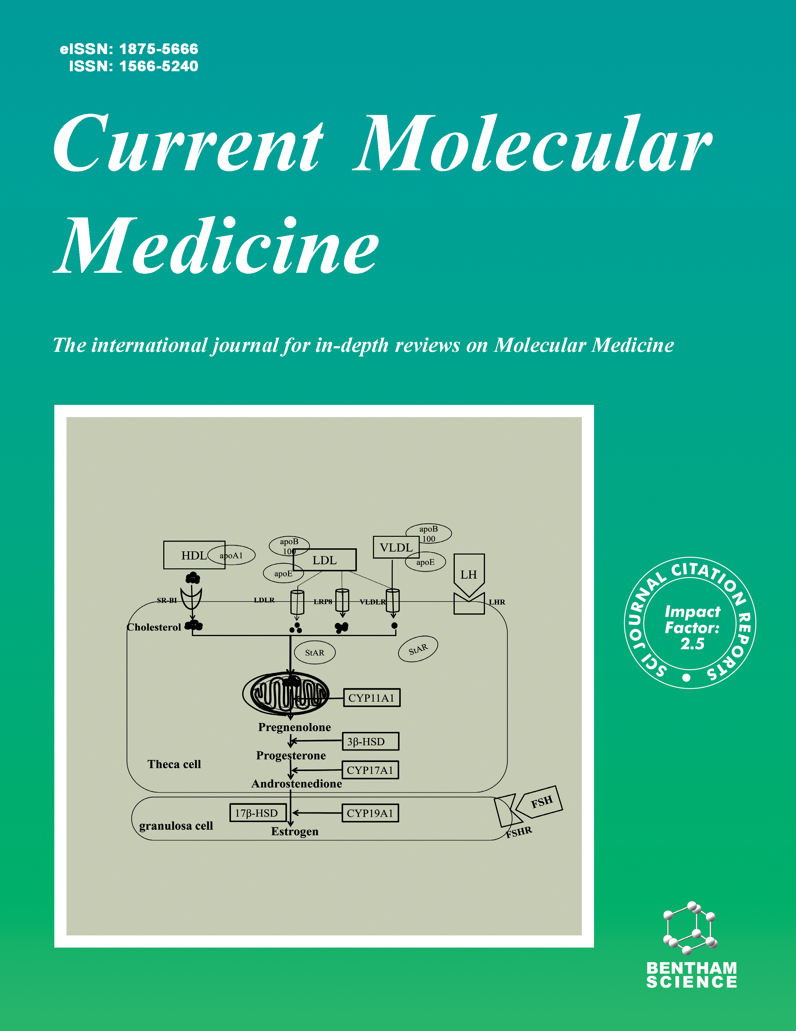
Current Molecular Medicine - Volume 14, Issue 6, 2014
Volume 14, Issue 6, 2014
-
-
Hepatocyte FRS2α is Essential for the Endocrine Fibroblast Growth Factor to Limit the Amplitude of Bile Acid Production Induced by Prandial Activity
More LessAuthors: C. Wang, C. Yang, J.Y.F. Chang, P. You, Y. Li, C. Jin, Y. Luo, X. Li, W.L. McKeehan and F. WangIn addition to being positively regulated by prandial activity, bile acid production is also negatively controlled by the endocrine fibroblast growth factor 19 (FGF19) or the mouse ortholog FGF15 from the ileum that represses hepatic cholesterol 7 α-hydroxylase (Cyp7a1) expression through activating FGF receptor four (FGFR4). However, how these two regulatory mechanisms interplay to control bile acid homeostasis in the body and the downstream pathways by which FGFR4 regulates Cyp7a1 expression are not fully understood. Here we report that hepatocyte FGFR substrate 2α (FRS2α), a scaffold protein essential for canonical FGFRs to activate the ERK and AKT pathways, was required for the regulation of bile acid production by the FGF15/19-FGFR4 signaling axis. This occurred through limiting the extent of increases in Cyp7a1 expression induced by prandial activity. Excess FGFR4 kinase activity reduced the amplitude of the increase whereas a lack of FGFR4 augmented the increase of Cyp7a1 expression in the liver. Ablation of Frs2α alleles in hepatocytes abrogated the regulation of Cyp7a1 expression by FGFR4. Together, the results demonstrate that FRS2α-mediated pathways are essential for the FGF15/FGF19-FGFR4 signaling axis to control bile acid homeostasis.
-
-
-
Activation of the Liver X Receptor Inhibits Th17 and Th1 Responses in Behcet’s Disease and Vogt-Koyanagi-Harada Disease
More LessBehcet’s disease (BD) and Vogt-Koyanagi-Harada (VKH) syndrome are two intraocular inflammatory diseases that are caused by an aberrant T lymphocyte response. Th17 cells, mainly producing the cytokine IL-17, and Th1 cells, characterized by the production of the index cytokine IFN-γ, are the CD4+ T lymphocyte subsets implicated in the pathogenesis of both BD and VKH. Suppressing the excessive response of these Th17 and Th1 cells has been reported to be an effective therapeutic approach to treat these patients and continuous efforts are being undertaken to find new methods to modulate the function of these cells. Evidence is emerging that the Liver X receptor (LXR) is an important regulator of inflammatory and immune responses and the study reported here was designed to investigate the role of LXR activation in BD and VKH. Here we demonstrate that the frequency of Th17 and Th1 cells along with the relevant cytokines IL-17, IFN-γ and corresponding transcriptional factors RORC, T-bet were all decreased following LXR activation by the agonist GW3965. LXR controlled the expression of inflammatory cytokines through an effect on NF-kappa B (NFκb) phosphorylation. Data from our study provide evidence for an association between a decreased LXR expression and disease activity in both BD and VKH, due to the fact that a lower LXR activation may result in an enhanced Th1 and Th17 immune response. Our study suggests that enhancing LXR activation may offer a potential therapeutic approach targeting aberrant immune responses by inhibiting Th1 and Th17 cell responses.
-
-
-
Apoptosis Induction by Ultrasound and Microbubble Mediated Drug Delivery and Gene Therapy
More LessAuthors: Z.-Y. Chen, Y.-X. Wang, Y.-Z. Zhao, F. Yang, J.-B. Liu, Y. Lin, J.-Y. Liao, Y.-Y. Liao and Q.-L. ZhouApoptosis induction provides a promising strategy for tumor gene therapy. Non-invasive ultrasound is a novel non-virus transfer method. In the field of cancer therapy, it has been found that ultrasound alone or together with microbubble represents an appealing, efficient and novel technique, which could deliver therapeutic gene or drug to specific organs or tissues in a simple and noninvasive way. Moreover, apoptosis induction mediated by the novel ultrasound-targeted microbubble destruction technique is safer and more effective than other methods, inactivating tumor cells, restraining cell proliferation and improving therapeutic effects of gene or chemotherapeutic drugs. In this paper, we reviewed apoptosis induction by ultrasound and microbubble mediated drug delivery and gene therapy in vitro and in vivo.
-
-
-
Knockdown of H19 Enhances Differentiation Capacity to Epidermis of Parthenogenetic Embryonic Stem Cells
More LessParthenogenetic embryonic stem (pES) cells are pluripotent stem cells derived from artificially activated oocytes without embryo destruction, thus eliciting less ethic concerns, and have been demonstrated promising for autologous stem cell therapy. However, pES cells could carry inappropriate imprinting such as relatively high expression of H19, a paternal imprinted gene, and may negatively influence their lineage differentiation. We show that knockdown of H19 by shRNA in mouse pES cells does not alter self-renewal and expression of genes associated with pluripotency. We find that down-regulation of H19 promotes differentiation of pES cells to epidermis. In addition, H19 depletion also facilitates differentiation of pES cells to cardiomyocytes and strong heart-like beating. Our data support the notion that reduction of H19 improves pES cell differentiation in the lineages of ectoderm and mesoderm, and provide further evidence suggesting that defective imprinting can be manipulated to allow potential application of pES cells for stem cell therapy.
-
-
-
Mesenchymal Stem Cells Regulate Cytoskeletal Dynamics and Promote Cancer Cell Invasion Through Low Dose Nitric Oxide
More LessBone marrow-derived mesenchymal stem cells (BMSCs) can be recruited to tumor sites and integrate into the stroma of tumors. When co-cultured with BMSCs, otherwise weakly metastatic nasopharyngeal carcinoma cells (NPC) showed improved metastatic ability. BMSCs in the tumor environment displayed the characteristics of macrophages. Nitric oxide produced by BMSCs in tumor environment could translocate caldesmon to podosome in Ca2+/calmodulin manner and promoted metastatic ability of NPC cells through invadopodia formation, with which the NPC cells degrade the extracellular matrix. Thus, we concluded that the BMSCs promoted cell migration and invasion through nitric oxide-induced paracrine signals.
-
-
-
Upregulation of Cytoskeleton Protein and Extracellular Matrix Protein Induced by Stromal-Derived Nitric Oxide Promotes Lung Cancer Invasion and Metastasis
More LessLung cancer commonly metastasizes to lymph nodes, brain and bones, which is the main cause of death. It is still a challenge to detect molecular biomarkers for early diagnosis and therapeutics of lung cancer. Our previous study found that bone marrow-derived stroma cells (BMSCs) under tumor microenvironment produced nitric oxide (NO), which was induced by inducible nitric oxide synthase (iNOS), and promoted invasion and metastasis of cancer cells by remodeling cytoskeleton. The aim of this study is to elucidate the relationship between the expressions of iNOS, cytoskeleton protein caldesmon, OPN, and clinical parameters especially the metastasis of lung cancer. We found that nitric oxide can remodel cytoskeleton and promoted the mobility of lung cancer cells. The expressions of iNOS, caldesmon, and OPN are closely correlated to metastasis of lung cancer. The intracranial metastatic tissue samples of lung cancer showed significantly higher expression of iNOS, caldesmon and OPN. A flow-cytometry analysis for peripheral blood of lung cancer patients showed increased EPCAM+/OPN+ cells in circulation of patients with bone metastasis compared to that of patients without metastasis, which is indicative of cancer circulating cells. The concentration of serum OPN was also positively related to the bone metastasis of lung cancer. Taken together, these results suggested that iNOS, caldesmon and OPN may work as biomarkers for metastasis of lung cancer.
-
-
-
The Mitochondrial Thioredoxin is Required for Liver Development in Zebrafish
More LessThioredoxins (Trxs) are a class of small molecular redox proteins that play an important role in scavenging abnormally accumulated reactive oxygen species (ROS). Thioredoxin 2 (Trx2) is one member of this family located in mitochondria. Trx2 protects cells from increased oxidative stress and has anti-apoptosis function. Knockout of Trx2 in mice led to early embryonic lethality. However, the essential role of Trx2 during embryogenesis remains unclear. To further investigate the role of Trx2 during embryonic development, we performed Trx2 knockdown in zebrafish and investigated the regulation role of Trx2 during embryonic development. Our results indicate that Trx2 had a high expression in early zebrafish embryos and its knockdown in zebrafish led to defective liver development mainly due to increased hepatic cell death. The increased ROS and the imbalance of members of the Bcl-2 family were involved in cell death induced by Trx2 suppression in zebrafish. The dysregulation of Bax, puma and Bcl-xl promoted the reduction of mitochondrial trans-membrane potential and the mitochondria membrane permeabilization (MMP), which initiated the mitochondrial apoptosis pathway. Additionally, we found that the increase of relocated GAPDH in mitochondria may be another factor responsible for the mitochondrial catastrophe.
-
-
-
Suppression of NF-κB Activation By Gentian Violet Promotes Osteoblastogenesis and Suppresses Osteoclastogenesis
More LessAuthors: M. Yamaguchi, T. Vikulina, J.L. Arbiser and M.N. WeitzmannSkeletal mass is regulated by the coordinated action of bone forming osteoblasts and bone resorbing osteoclasts. Accelerated rates of bone resorption relative to bone formation lead to net bone loss and the development of osteoporosis, a devastating disease that predisposes the skeleton to fractures. Bone fractures are associated with significant morbidity and in the case of hip fractures, high mortality. Gentian violet (GV), a cationic triphenylmethane dye, has long been used as an antifungal and antibacterial agent and is presently under investigation as a potential chemotherapeutic and antiangiogenic agent. However, effects on bone cells have not been previously reported and the mechanisms of action of GV, are poorly understood. In this study we show that GV suppresses receptor activator of NF-κB ligand (RANKL)-induced differentiation of RAW264.7 osteoclast precursors into mature osteoclasts, but paradoxically stimulates the differentiation of MC3T3 cells into mineralizing osteoblasts. These actions stem from the capacity of GV to suppress activation of the nuclear factor kappa B (NF-κB) signal transduction pathway that is required for osteoclastogenesis, but inhibitory to osteoblast differentiation and activity. Our data reveal that GV is an inhibitor of NF-κB activation and may hold promise for modulation of bone turnover to promote a balance between bone formation and bone resorption, favorable to gain of bone mass.
-
-
-
A Mixed Anti-Inflammatory and Pro-Inflammatory Response Associated with a High Dose of Corticosteroids
More LessAuthors: P. Dandona, H. Ghanim, C.L. Sia, K. Green, S. Abuaysheh, S. Dhindsa, A. Chaudhuri and A. MakdissiObjective: Hydrocortisone, at a low dose (100 mg), induces an anti-inflammatory response including inducing IkBα and suppressing intranuclear NFκB and AP-1 binding and the expression of pro-inflammatory mediators like MMPs. We have now investigated the effect of a high dose of hydrocortisone (300mg=60 mg prednisolone) on NFκB binding and the expression of TLRs, the mediators of TLR signal transduction, MyD88 and TRIF and HMG-B1. Design and Subjects: A 300mg of hydrocortisone or saline was injected intravenously in ten normal subjects during 2 separate visits, in a randomized crossover study. Blood samples were obtained at 0, 1, 4, 6 and 24h after the injection and mononuclear cells (MNC) were prepared. Results: There was a significant increase in glucose (from 92±4 to 116±6 mg/dl), insulin (from 4.5±0.7 to 5.3±0.8 mU/ml) and FFA concentrations (from 0.38±0.1 to 0.80±0.15mM) following the administration of hydrocortisone compared to placebo treatment. While NFκB binding and the mRNA expression of MyD88, TRIF, chemokines and chemokine receptors were suppressed significantly in MNC, there was a paradoxical increase in the mRNA expression of TLR 2, 5 and 9 and HMG-B1 was increased by 103±24%, 107±19%, 56±13% and 58±12% above the baseline, respectively in the MNC. Plasma concentrations of HMG-B1 and MMP-9 increased by 37±12% and 125±22%, respectively, while TNF-α concentrations fell by 27±9%. Conclusion: While this high dose of hydrocortisone exerts a powerful anti-inflammatory effect, it also exerts certain proinflammatory effects mainly on TLRs expression. The known pro-inflammatory effects of glucose and FFAs may have contributed to these effects. These paradoxical pro-inflammatory effects may account for the inability of these drugs to show benefit in clinical trials of septicemia and other severe pro-inflammatory states and might contribute to some of the side effects of corticosteroids use.
-
Volumes & issues
-
Volume 25 (2025)
-
Volume 24 (2024)
-
Volume 23 (2023)
-
Volume 22 (2022)
-
Volume 21 (2021)
-
Volume 20 (2020)
-
Volume 19 (2019)
-
Volume 18 (2018)
-
Volume 17 (2017)
-
Volume 16 (2016)
-
Volume 15 (2015)
-
Volume 14 (2014)
-
Volume 13 (2013)
-
Volume 12 (2012)
-
Volume 11 (2011)
-
Volume 10 (2010)
-
Volume 9 (2009)
-
Volume 8 (2008)
-
Volume 7 (2007)
-
Volume 6 (2006)
-
Volume 5 (2005)
-
Volume 4 (2004)
-
Volume 3 (2003)
-
Volume 2 (2002)
-
Volume 1 (2001)
Most Read This Month


