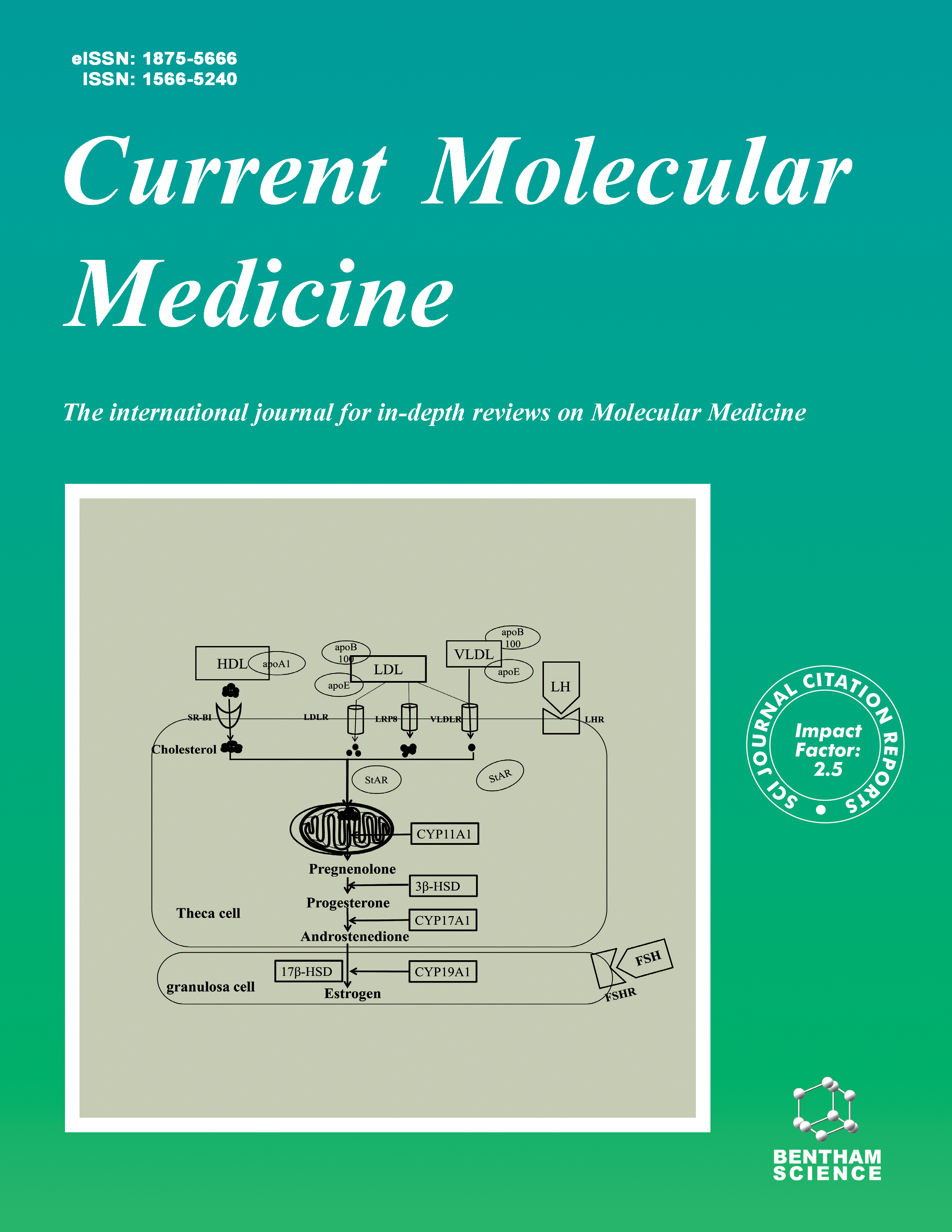
Current Molecular Medicine - Volume 8, Issue 3, 2008
Volume 8, Issue 3, 2008
-
-
The Stress Rheostat: An Interplay Between the Unfolded Protein Response (UPR) and Autophagy in Neurodegeneration
More LessAuthors: Soledad Matus, Fernanda Lisbona, Mauricio Torres, Cristian Leon, Peter Thielen and Claudio HetzThe unfolded protein response (UPR) is a conserved adaptive reaction that increases cell survival under conditions of endoplasmic reticulum (ER) stress. The UPR controls diverse processes such as protein folding, secretion, ER biogenesis, protein quality control and macroautophagy. Occurrence of chronic ER stress has been extensively described in neurodegenerative conditions linked to protein misfolding and aggregation, including Amyotrophic lateral sclerosis, Prion-related disorders, and conditions such as Parkinson's, Huntington's, and Alzheimer's disease. Strong correlations are observed between disease progression, accumulation of protein aggregates, and induction of the UPR in animal and in vitro models of neurodegeneration. In addition, the first reports are available describing the engagement of ER stress responses in brain postmortem samples from human patients. Despite such findings, the role of the UPR in the central nervous system has not been addressed directly and its contribution to neurodegeneration remains speculative. Recently, however, pharmacological manipulation of ER stress and autophagy - a stress pathway modulated by the UPR - using chemical chaperones and autophagy activators has shown therapeutic benefits by attenuating protein misfolding in models of neurodegenerative disease. The most recent evidence addressing the role of the UPR and ER stress in neurodegenerative disorders is reviewed here, along with therapeutic strategies to alleviate ER stress in a disease context.
-
-
-
Programmed Cell Death Mechanisms in Neurological Disease
More LessProgrammed cell death (pcd) is a form of cell death in which the cell plays an active role in its own demise. Pcd plays a critical role in the development of the nervous system, as well as in its response to insult. Both anti-pcd and pro-pcd modulators play prominent roles in development and disease, including neurodegeneration, cancer, and ischemic vascular disease, among others. Over 100,000 published studies on one form of programmed cell death—apoptosis—have appeared, but recent studies from multiple laboratories suggest the existence of non-apoptotic forms of programmed cell death, such as autophagic programmed cell death. In addition, there appear to be programmatic cell deaths that do not fit the criteria for either apoptosis or autophagic cell death, arguing that additional programs may also be available to cells. Constructing a mechanistic taxonomy of all forms of pcd—based on inhibitors, activators, and identified biochemical pathways involved in each form of pcd—should offer new insight into cell deaths associated with various disease states, and ultimately offer new therapeutic approaches.
-
-
-
Cell Death by Necrosis, a Regulated Way to Go
More LessAuthors: Mauricio Henriquez, Ricardo Armisen, Andres Stutzin and Andrew F.G. QuestApoptosis is a programmed form of cell death with well-defined morphological traits that are often associated with activation of caspases. More recently evidence has become available demonstrating that upon caspase inhibition alternative programs of cell death are executed, including ones with features characteristic of necrosis. These findings have changed our view of necrosis as a passive and essentially accidental form of cell death to that of an active, regulated and controllable process. Also necrosis has now been observed in parallel with, rather than as an alternative pathway to, apoptosis. Thus, cell death responses are extremely flexible despite being programmed. In this review, some of the hallmarks of different programmed cell death modes have been highlighted before focusing the discussion on necrosis. Obligatory events associated with this form of cell death include uncompensated cell swelling and related changes at the plasma membrane. In this context, representatives of the transient receptor channel family and their regulation are discussed. Also mechanisms that lead to execution of the necrotic cell death program are highlighted. Emphasis is laid on summarizing our understanding of events that permit switching between cell death modes and how they connect to necrosis. Finally, potential implications for the treatment of some disease states are mentioned.
-
-
-
Molecular Mechanisms and Pathophysiology of Necrotic Cell Death
More LessAuthors: Nele Vanlangenakker, Tom V. Berghe, Dmitri V. Krysko, Nele Festjens and Peter VandenabeeleNecrotic cell death has long been considered an accidental and uncontrolled mode of cell death. But recently it has become clear that necrosis is a molecularly regulated event that is associated with pathologies such as ischemia-reperfusion (IR) injury, neurodegeneration and pathogen infection. The serine/threonine kinase receptor-interacting protein 1 (RIP1) plays a crucial role during the initiation of necrosis induced by ligand- receptor interactions. On the other hand, ATP depletion is an initiating factor in ischemia-induced necrotic cell death. Common players in necrotic cell death irrespective of the stimulus are calcium and reactive oxygen species (ROS). During necrosis, elevated cytosolic calcium levels typically lead to mitochondrial calcium overload, bioenergetics effects, and activation of proteases and phospholipases. ROS initiates damage to lipids, proteins and DNA and consequently results in mitochondrial dysfunction, ion balance deregulation and loss of membrane integrity. Membrane destabilization during necrosis is also mediated by other factors, such as acidsphingomyelinase (ASM), phospholipase A2 (PLA2) and calpains. Furthermore, necrotic cells release immunomodulatory factors that lead to recognition and engulfment by phagocytes and the subsequent immunological response. The knowledge of the molecular mechanisms involved in necrosis has contributed to our understanding of necrosis-associated pathologies. In this review we will focus on the intracellular and intercellular signaling events in necrosis induced by different stimuli, such as oxidative stress, cytokines and pathogenassociated molecular patterns (PAMPs), which can be linked to several pathologies such as stroke, cardiac failure, neurodegenerative diseases, and infections.
-
-
-
Molecular Genetics and Biomarkers of Polyglutamine Diseases
More LessPolyglutamine diseases are hereditary neurodegenerative disorders caused by an abnormal expansion of a trinucleotide CAG repeat, which encodes a polyglutamine tract. To date, nine polyglutamine diseases are known: Huntington's disease (HD), spinal and bulbar muscular atrophy (SBMA), dentatorubralpallidoluysian atrophy (DRPLA) and six forms of spinocerebellar ataxia (SCA). The diseases are inherited in an autosomal dominant fashion except for SBMA, which shows an X-linked pattern of inheritance. Although the causative gene varies with each disorder, polyglutamine diseases share salient genetic features as well as molecular pathogenesis. CAG repeat size correlates well with the age of onset in each disease, shows both somatic and germline instability, and has a strong tendency to further expand in successive generations. Aggregation of the mutant protein followed by the disruption of cellular functions, such as transcription and axonal transport, has been implicated in the etiology of neurodegeneration in polyglutamine diseases. Although animal studies have provided promising therapeutic strategies for polyglutamine diseases, it remains difficult to translate these disease-modifying therapies to the clinic. To optimize “proof of concept”, the process for testing candidate therapies in humans, it is of importance to identify biomarkers which can be used as surrogate endpoints in clinical trials for polyglutamine diseases.
-
-
-
Cutaneous Melanoma: Fishing with Chips
More LessAuthors: Sandeep Nambiar, Alireza Mirmohammadsadegh and Ulrich R. HenggeDNA microarray technology is a versatile platform that allows rapid genetic analysis to take place on a genome-wide scale and has revolutionized the way cancers are studied. This platform has enabled researchers to characterize mechanisms central to tumorigenesis and understand important molecular events in the multi-step tumor progression model of cutaneous melanoma and other cancers. In melanoma, multiple global gene expression profiling studies using various DNA microarray platforms and various experimental designs have been performed. Each study has been able to capture and characterize either the involvement of a novel pathway or a novel cause-effect-relationship. The use of microarrays to define subclasses, to identify differentially regulated genes within a mutational context to analyze epigenetically regulated genes has resulted in an unprecedented understanding of the biology of cutaneous melanoma that may lead to more accurate diagnosis, more comprehensive prognosis, prediction and more effective therapeutic interventions. Related DNA microarray platforms like array-comparative genomic hybridization (CGH) have also been instrumental to identify many non-random chromosomal alterations; however, studies identifying validated targets as a result of CGH are limited. Thus, there exists significant opportunity to discover novel melanoma genes and translate such discoveries into meaningful clinical endpoints. In this review, we focus on various DNA microarray-based studies performed in cutaneous melanoma and summarize our current understanding of the genetics and biology of melanoma progression derived from accumulating genomic information.
-
Volumes & issues
-
Volume 25 (2025)
-
Volume 24 (2024)
-
Volume 23 (2023)
-
Volume 22 (2022)
-
Volume 21 (2021)
-
Volume 20 (2020)
-
Volume 19 (2019)
-
Volume 18 (2018)
-
Volume 17 (2017)
-
Volume 16 (2016)
-
Volume 15 (2015)
-
Volume 14 (2014)
-
Volume 13 (2013)
-
Volume 12 (2012)
-
Volume 11 (2011)
-
Volume 10 (2010)
-
Volume 9 (2009)
-
Volume 8 (2008)
-
Volume 7 (2007)
-
Volume 6 (2006)
-
Volume 5 (2005)
-
Volume 4 (2004)
-
Volume 3 (2003)
-
Volume 2 (2002)
-
Volume 1 (2001)
Most Read This Month


