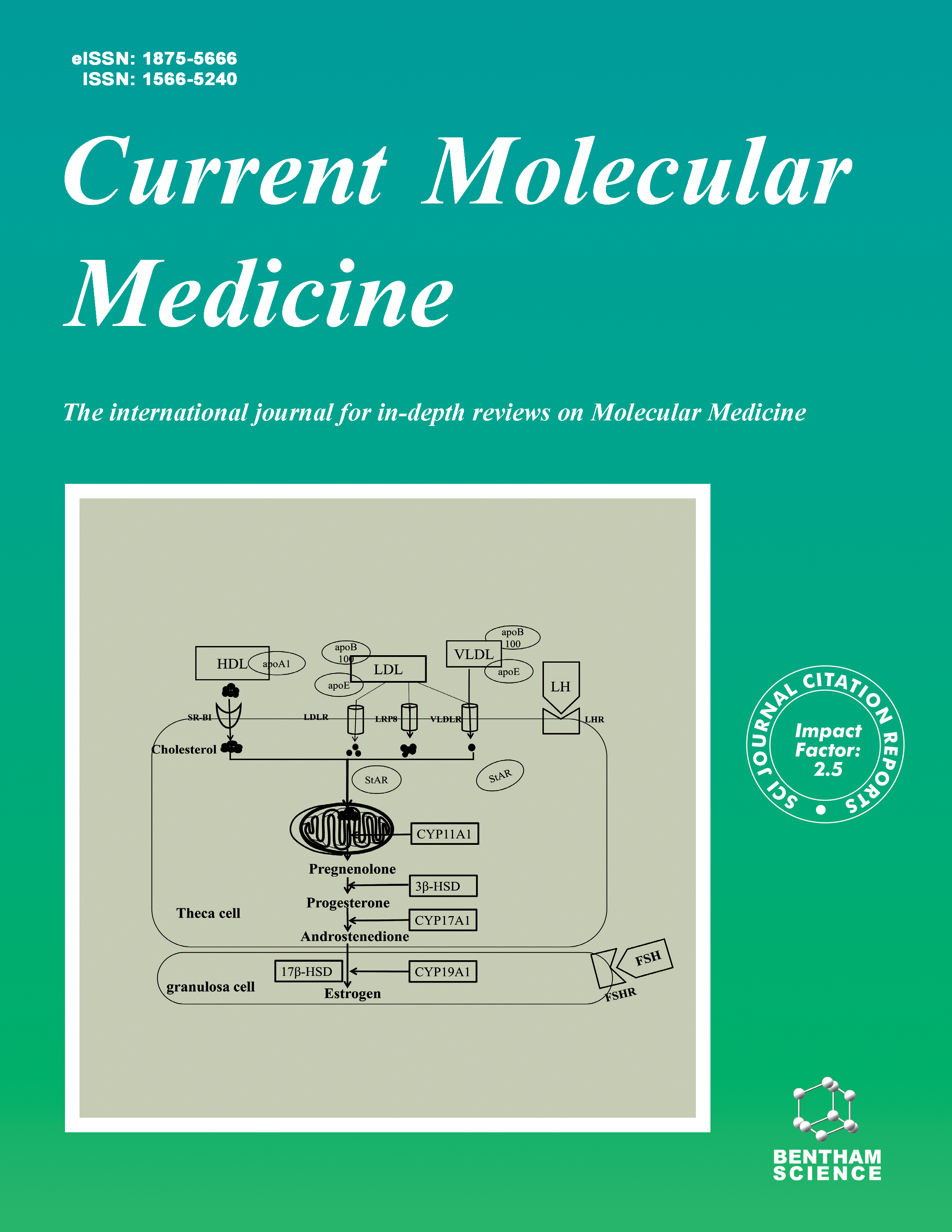
Current Molecular Medicine - Volume 23, Issue 6, 2023
Volume 23, Issue 6, 2023
-
-
Glaucoma: Biological Mechanism and its Clinical Translation
More LessGlaucoma is a common cause of visual loss and irreversible blindness, affecting visual and life quality. Various mechanisms are involved in retinal ganglion cell (RGC) apoptosis and functional and structural loss in the visual system. The prevalence of glaucoma has increased in several countries. However, its early diagnosis has contributed to prompt attention. Molecular and cellular biological mechanisms are important for understanding the pathological process of glaucoma and new therapies. Thus, this review discusses the factors involved in glaucoma, from basic science to cellular and molecular events (e.g., mitochondrial dysfunction, endoplasmic reticulum stress, glutamate excitotoxicity, the cholinergic system, and genetic and epigenetic factors), which in recent years have been included in the development of new therapies, management, and diagnosis of this disease.
-
-
-
Current Status of Alzheimer’s Disease and Pathological Mechanisms Investigating the Therapeutic Molecular Targets
More LessAuthors: Shivani Bagga and Manish KumarAlzheimer's disease (AD) is a psychological, biological, or developmental disorder that affects basic mental functioning. AD is generally affiliated with marked discomfort and impaired social, professional, or other crucial aspects of life. AD is predominant worldwide, but a disparity in prevalence is observed amongst nations. Around 3/4 of people with Alzheimer's disease are from underdeveloped nations, which receive only 1/10th of global mental health resources. Residents of each community and age category share their presence in the overall load of AD. AD is a multifactorial disease impacted by numerous environmental, genetic, and endogenous elements. Heteromorphic interactive downstream cascades, networks, and molecular mechanisms (inflammation and immune network, cholinergic deficit, lipid transit, endocytosis, excitotoxicity, oxidative stress, amyloid and tau pathology, energy metabolism, neuron and synapse loss, and cell death) have been isolated, imparting a non-dissociative contribution in pathogenesis of AD. In the CNS, the structural organization of cholinergic neurons can give a novel insight into the mechanism of new learning. The alleviation of central cholinergic transposal following destruction in the basal forebrain cholinergic neurons precipitates a decline in neurocognitive symptoms visible in AD patients. The brain of patients suffering from AD exhibits plaques of aggregated amyloid-β and neurofibrillary tangles containing hyperphosphorylated tau protein. Amyloid-β triggers cholinergic loss by modulation of calcium and generation of cell-damaging molecules such as nitric oxide and reactive oxygen species intermediates. The present review focuses on the pathogenic mechanisms related to stages, diagnosis, and therapeutic approaches involved in AD.
-
-
-
Advances in Exosomal microRNAs and Proteins in Ovarian Cancer Diagnosis, Prognosis, and Treatment
More LessAuthors: Tiansheng Qin, Fan Chen, Jiaojiao Zhu, Yaoyao Ding and Qianqian ZhangLate diagnosis, postoperative recurrence, and chemotherapy resistance are the main causes of the high mortality rate in ovarian cancer (OC). Understanding the molecular mechanisms in the pathogenesis and progression of OC may contribute to discovering new tumor biomarkers and therapeutic targets for OC. Exosomes are small extracellular vesicles derived from different types of cells that carry cargos, including nucleic acids, proteins, and lipids, and are pivotal mediators of intercellular communication in the tumor microenvironment. There is emerging evidence that exosomal proteins and nucleic acids play pivotal roles in facilitating the progression and drug resistance of OC. Identification of these factors may aid in the future diagnosis of OC. Furthermore, they also have promising value as OC therapeutic targets that can improve the prognosis. In the current review, we summarize the progress of exosomal research in OC, especially highlighting the most updated roles of exosomal microRNAs and proteins in the diagnosis, prognosis, therapy, and drug resistance of OC in order to facilitate future studies in this area.
-
-
-
CTLA-4: As an Immunosuppressive Immune Checkpoint in Breast Cancer
More LessBreast cancer (BC) is one of the prevalent diseases and causes of death in women, and its incidence rate is increasing in numerous developed and developing countries. The common approach to BC therapy is surgery, followed by radiation therapy or chemotherapy, which doesn't lead to acceptable outcomes in many patients. Therefore, developing innovative strategies for treating BC is essential for the most effective therapy. The immunotherapy of BC is a promising and attractive strategy that can increase the immune system's capacity to recognize and kill the tumor cells, inhibit the recurrence of the tumors, and develop new metastatic sites. The blockade of immune checkpoints is the most attractive and promising strategy for cancer immunotherapy. The cytotoxic T lymphocyte-associated antigen-4 (CTLA-4) is a cellsurface glycoprotein expressed by stimulated T cells and has pivotal roles in cell cycle modulation, cytokine generation, and regulation of T cell proliferation. Currently, anti- CTLA-4 agents such as monoclonal antibodies (Ipilimumab and tremelimumab) are broadly applied as therapeutic agents in clinical studies of different cancers. The anti- CTLA-4 antibodies, alone or combined with other therapeutic agents, remarkably increased the tumor-suppressive effects of the immune system and improved the prognosis of cancer. The immune checkpoint inhibitors may represent promising options for BC treatment as in monotherapy or in combination with other conventional treatments. In this review, we discuss the role of CTLA-4 and its therapeutic potential by inhibitors of immune checkpoints in BC therapeutics.
-
-
-
Crocetin Suppresses Uterine Ischemia/Reperfusion-induced Inflammation and Apoptosis through the Nrf-2/HO-1 Pathway
More LessBackground: Uterine ischemia/reperfusion (I/R) injury often occurs during many complex surgical procedures, such as uterus transplantation, cesarean, and myomectomy, which may lead to the loss of uterine function and failure of the operation. Crocetin (CRO), as one of the major active constituents from saffron extract, shows protective effects against reactive oxygen species, inflammation, and apoptosis. However, the role of CRO in protecting the uterus against I/R-induced injury has never been investigated. This study aims to clarify the protective role of CRO against I/R injury and the underlying mechanisms. Materials and Methods: Sprague-Dawley rats were randomly divided into five groups: the control group, I/R group, 20 mg/kg CRO-treated I/R group, 40 mg/kg CRO-treated I/R group, and 80 mg/kg CRO-treated I/R group. Rats were given daily gavages with different doses of CRO or vehicle for five consecutive days. The rat uterine I/R model was created by routine method with 1h ischemia and 3h reperfusion. The serum and uterine tissues were collected, the changes in malondialdehyde (MDA) level and superoxide dismutase (SOD) activity, the mRNA and protein levels of interleukin (IL)-1β, IL-6, tumor necrosis factor (TNF)-α and IL-10, the protein levels of B-cell chronic lymphocytic leukemia/lymphoma (Bcl)-2, Bcl-2-associated X protein (Bax), caspase-3, nuclear factor erythroid 2-related factor (Nrf)-2, and heme oxygenase (HO)-1, were measured. The histological changes were examined by HE staining. The number of apoptotic cells was analyzed by flow cytometry. Results: Uterine I/R significantly induced MDA level, suppressed SOD activity, upregulated levels of pro-inflammatory cytokines, down-regulated level of the antiinflammatory cytokine, induced caspase-3-dependent apoptosis, activated the protein expression of Nrf-2 and HO-1, and caused uterine damage. However, pre-administration of CRO effectively reversed I/R-induced above changes and further enhanced Nrf-2/HO- 1 activation in a dose-dependent manner. Conclusion: Pre-administration of CRO effectively alleviates I/R-induced oxidative stress, inflammation, apoptosis, and tissue injury probably through activating the Nrf- 2/HO-1 pathway, suggesting a protective role of CRO in I/R-induced uterus injury.
-
-
-
Verapamil Regulates the Macrophage Immunity to Mycobacterium tuberculosis through NF-ΚB Signaling
More LessAuthors: Wenping Gong, Ruina Cui, Lele Song, Yourong Yang, Junxian Zhang, Yan Liang, Xuejuan Bai, Jie Wang, Lan Wang, Xueqiong Wu and Weiguo ZhaoBackground: Verapamil enhances the sensitivity of Mycobacterium tuberculosis to anti-tuberculosis (TB) drugs, promotes the macrophage anti-TB ability, and reduces drug resistance, but its mechanism is unclear. Herein, we have investigated the effect of verapamil on cytokine expression in mouse peritoneal macrophages. Methods: Macrophages from mice infected with M. tuberculosis or S. aureus were cultured with verapamil, the cytokines were detected by enzyme-linked immunosorbent assay, and the RNA was measured with quantitative real-time polymerase chain reaction and agarose gel electrophoresis. The intracellular calcium signaling was measured by confocal microscopy. Results: Significantly higher levels of NF-ΚB, IL-12, TNF-α, and IL-1β were observed after TB infection. The levels of NF-ΚB and IL-12 increased when verapamil concentration was less than 50 μg/ml, but decreased when verapamil concentration was greater than 50μg/ml. With the increase in verapamil concentration, TNF-α and IL-1β expressed by macrophages decreased. The L-type calcium channel transcription significantly increased in M. tuberculosis rather than S. aureus-infected macrophages. Furthermore, during bacillus Calmette-Guerin (BCG) infection, verapamil stimulated a sharp peak in calcium concentration in macrophages, while calcium concentration increased mildly and decreased smoothly over time in the absence of verapamil. Conclusion: Verapamil enhanced macrophage immunity via the NF-ΚB pathway, and its effects on cytokine expression may be achieved by its regulation of intracellular calcium signaling.
-
-
-
Ursodeoxycholic Acid (UDCA) Reduces Hepatocyte Apoptosis by Inhibiting Farnesoid X Receptor (FXR) in Hemorrhagic Shock (HS)
More LessAuthors: Lu Wang, Xi Rui, Huai-Wu He, Xiang Zhou and Yun LongBackground: Hemorrhagic shock (HS) is the most common cause of potentially preventable death after traumatic injury. Acute liver injury is an important manifestation of HS. Apoptosis plays an important role in liver injury. Farnesoid X receptor (FXR) can alleviate liver injury. This study aimed to examine the effects of ursodeoxycholic acid (UDCA) on hepatocyte apoptosis in HS and its relationship with the FXR pathway. Methods: Mice were randomly divided into 4 groups: sham group, HS group, HS + UDCA group, and FXR (-) + HS + UDCA group. There were 6 mice in each group. As to the model of HS, MAP of 40 ± 5 mmHg was maintained for 1 hour. As to UDCA intervention, UDCA (300mg/kg) was given nasally. Real-time RT-PCR and Western blotting were used to detect changes in the expression level of Caspase-3, Bax, LC3133;, LC3133;¡, Bcl-2, and Beclin-1 in the liver. TUNEL assay was used to detect changes in hepatocyte apoptosis. Results: The expression level of Caspase-3 and Bax in the liver decreased significantly after treatment with UDCA under HS conditions. The expression level of LC3133;, LC3133;¡, Bcl-2, and Beclin-1 in the liver increased significantly after treatment with UDCA under HS conditions. TUNEL positive percentage of liver decreased significantly after treatment with UDCA under HS conditions. In the case of FXR (-), the influence of UDCA was inhibited. Conclusion: These results indicated that UDCA could reduce hepatocyte apoptosis during HS through the FXR pathway.
-
-
-
Transcriptome Profiling of Cisplatin Resistance in Triple-negative Breast Cancer: New Insight into the Role of PI3k/Akt Pathway
More LessAuthors: Maryam Memar, Touraj Farazmandfar, Amir Sabaghian, Majid Shahbazi and Masoud GolalipourBackground: Aggressive nature of triple negative breast cancer (TNBC) is associated with poor prognosis compared with other breast cancer types. Current guidelines recommend the use of Cisplatin for the management of TNBC. However, the development of resistance to cisplatin is the primary cause of chemotherapy failure. Objective: In the present study, we aimed to develop a stable cisplatin-resistant TNBC cell line to investigate the key pathways and genes involved in cisplatin-resistant TNBC. Methods: The MDA-MB-231 cell was exposed to different concentrations of cisplatin. After 33 generations, cells showed a resistant phenotype. Then, RNA-sequencing analysis was performed in cisplatin-resistant and parent cell lines. The RNA-sequencing data was verified by quantitative PCR (qPCR). Results: The IC50 of the resistant cell increased to 10-fold of a parental cell (p<0.001). Also, cisplatin-resistant cells show cross-resistance to other drugs, including 5- fluorouracil, paclitaxel, and doxorubicin. Resistant cells demonstrated reduced drug accumulation compared to the parental cells. Results showed there were 116 differentially expression genes (DEGs) (p<0.01). Gene ontology analysis revealed that the DEGs have several molecular functions, including binding and transporter activity. Functional annotation showed that the DEGs were enriched in the drug resistancerelated pathways, especially the PI3K-Akt signaling pathway. The most important genes identified in the protein-protein interaction network were heme oxygenase 1 (HMOX1) and TIMP metallopeptidase inhibitor 3 (TIMP3). Conclusion: We have identified several pathways and DEGs associated with the PI3KAkt pathway, which provides new insights into the mechanism of cisplatin resistance, and potential drug targets in TNBC.
-
-
-
Dexmedetomidine Attenuates Spinal Cord Ischemia-reperfusion Injury in Rabbits by Decreasing Oxidation and Apoptosis
More LessAuthors: Bingbing Liu, Yatong Liang, Weihua Huang, Hui Zhang, Daiwei Zhou and Xiaoshan XiaoBackground: In brain ischemia, dexmedetomidine (DEX) prevents glutamate and norepinephrine changes, increases nerve conduction, and prevents apoptosis, but the mechanisms are poorly understood. Objective: This study aimed at examining the protective effect and function of DEX on spinal cord ischemia-reperfusion injury (SCIRI) and whether the effect is mediated by oxidative stress and apoptosis (with the involvement of Bcl-2, Bax, mitochondria, and Caspase-3). Methods: Rabbits were randomly divided into the sham group, infusion/reperfusion (I/R) group, and DEX+I/R group. SCIRI was induced by occluding the aorta just caudal to the left renal artery for 40 min, followed by reperfusion. DEX was continuously administered for 60 min before clamping. The animals were evaluated for neuronal functions. Spinal cord tissues were examined for SOD activity and MDA content. Bcl-2, Bax, and Caspase-3 expressions were detected by western blotting. TUNEL staining was used for apoptosis. Results: With the extension of reperfusion time, the hind limbs’ neurological function in the DEX+I/R group gradually improved, but it became worse in the I/R group (all P<0.05 vs. the other time points within the same groups). Compared with I/R, DEX decreased MDA and increased SOD (P<0.01), upregulated Bcl-2 protein expression (P<0.05), downregulated Bax expression (P<0.05), decreased caspase-3 expression (P<0.05), prevented histological changes in neurons, and decreased the apoptotic index of the TUNEL labeling (P<0.05). Conclusion: DEX could attenuate SCIRI in rabbits by improving the oxidative stress status, regulating the expression of apoptosis-related proteins, and decreasing neuronal apoptosis.
-
-
-
Bio-Membrane SELEX as a New Approach for Selecting ss-DNA Aptamers that Bind to the Hydatid Cyst Laminated Layer
More LessBackground: Hydatid cyst (HC) is the larval stage of the canine intestinal tapeworm (cestode), Echinococcus granulosus. In addition to the high global economic cost of livestock farming, the infection can lead to dangerous problems for human health. Therefore, research into new diagnosis and treatment approaches is valuable. This study is set out to explore aptamers that bind to HC antigens. Methods: The similarity between HC genotype in sheep and humans was that sheep HCs were collected and used as a biological membrane for aptamer selection. Four Bio- Membrane SELEX rounds were conducted, and ssDNA aptamers were selected. Selected aptamers' affinity and specificity to the laminated layer antigens were evaluated using membrane staining by fluorescein primer as a probe. Biotinylated primer was used as a probe for aptahistochemistry and dot blot techniques. Subsequently, cloning and plasmid extraction was conducted. The affinity and specificity of sequenced aptamers were examined with the dot blot method. Results: Selected aptamers reacted with HC wall in aptahistochemistry, aptahistofluorescent, and dot blot experiments. Following cloning and sequencing, 20 sequences were achieved. A strong reaction between HC total antigens and sequenced aptamers has emerged in the dot blot method. Conclusion: We propose a novel method to determine specific aptamers in this investigation. Bio-Membrane SELEX could be assumed as a practical and sensitive method for aptamer selection. Selected aptamers in this study possibly may be used for specific HC antigens detection.
-
Volumes & issues
-
Volume 25 (2025)
-
Volume 24 (2024)
-
Volume 23 (2023)
-
Volume 22 (2022)
-
Volume 21 (2021)
-
Volume 20 (2020)
-
Volume 19 (2019)
-
Volume 18 (2018)
-
Volume 17 (2017)
-
Volume 16 (2016)
-
Volume 15 (2015)
-
Volume 14 (2014)
-
Volume 13 (2013)
-
Volume 12 (2012)
-
Volume 11 (2011)
-
Volume 10 (2010)
-
Volume 9 (2009)
-
Volume 8 (2008)
-
Volume 7 (2007)
-
Volume 6 (2006)
-
Volume 5 (2005)
-
Volume 4 (2004)
-
Volume 3 (2003)
-
Volume 2 (2002)
-
Volume 1 (2001)
Most Read This Month


