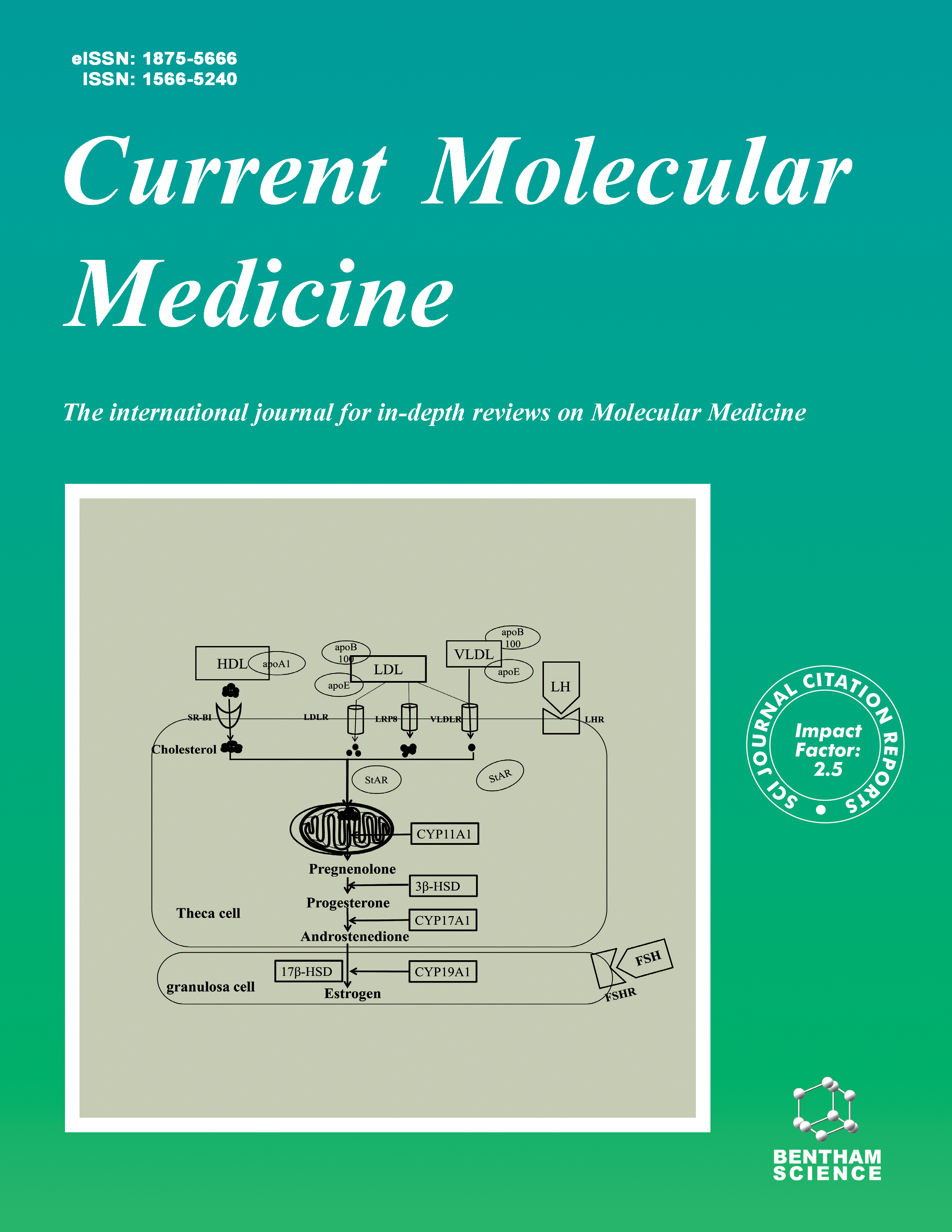
Current Molecular Medicine - Volume 20, Issue 9, 2020
Volume 20, Issue 9, 2020
-
-
The Roles of microRNAs in Multidrug-Resistance Mechanisms in Gastric Cancer
More LessAuthors: Xi Zeng, Hao-Ying Wang, Su-Yang Bai, Ke Pu, Yu-Ping Wang and Yong-Ning ZhouMultidrug resistance (MDR) is one of the most significant reasons for the chemotherapeutics failure in gastric cancer. Although accumulating investigations and researches have been made to elucidate the mechanisms of multidrug resistance, the detail is far from completely understood. The importance of microRNAs in cancer chemotherapeutic resistance has been demonstrated recently, which provides a new strategy to overcome multidrug resistance. The different mechanisms are related to the phenomena of MDR itself and the roles of miRNAs in these multi-mechanisms by which MDR is acquired. In turn, the aim of this review was to summarize recent publications of microRNAs in regulating MDR in gastric cancer, thereby potentially developing as targeted therapies. Further unraveling the roles of microRNAs in MDR mechanisms including the ATP-binding cassette (ABC) transporter family, autophagy induction, cancer stem cell regulation, hypoxia induction, DNA damage and repair, epigenetic regulation, and exosomes in gastric cancer will be helpful for us to win the battle against it.
-
-
-
Hyaluronic Acid and Regenerative Medicine: New Insights into the Stroke Therapy
More LessStroke is known as one of the very important public health problems that are related to societal burden and tremendous economic losses. It has been shown that there are few therapeutic approaches for the treatment of this disease. In this regard, the present therapeutic platforms aim to obtain neuroprotection, reperfusion, and neuro recovery. Among these therapies, regenerative medicine-based therapies have appeared as new ways of stroke therapy. Hyaluronic acid (HA) is a new candidate, which could be applied as a regenerative medicine-based therapy in the treatment of stroke. HA is a glycosaminoglycan composed of disaccharide repeating elements (N-acetyl-Dglucosamine and D-glucuronic acid). Multiple lines of evidence demonstrated that HA has critical roles in normal tissues. It can be a key player in different physiological and pathophysiological conditions such as water homeostasis, multiple drug resistance, inflammatory processes, tumorigenesis, angiogenesis, and changed viscoelasticity of the extracellular matrix. HA has very important physicochemical properties i.e., availability of reactive functional groups and its solubility, which make it a biocompatible material for application in regenerative medicine. Given that HAbased bioscaffolds and biomaterials do not induce inflammation or allergies and are hydrophilic, they are used as soft tissue fillers and injectable dermal fillers. Several studies indicated that HA could be employed as a new therapeutic candidate in the treatment of stroke. These studies documented that HA and HA-based therapies exert their pharmacological effects via affecting stroke-related processes. Herein, we summarized the role of the extracellular matrix in stroke pathogenesis. Moreover, we highlighted the HA-based therapies for the treatment of stroke.
-
-
-
Cholesterol: A Prelate in Cell Nucleus and its Serendipity
More LessAuthors: Nimisha Saxena and Nimai C. ChandraCholesterol is a chameleon bio-molecule in cellular multiplex. It acts as a prelate in almost every cellular compartment with its site specific characteristics viz. regulation of structural veracity and scaffold fluidity of bio-membranes, insulation of electrical transmission in nerves, controlling of genes by making steroid endocrines, acting as precursors of metabolic regulators and many more with its emerging prophecy in the cell nucleus to drive new cell formation. Besides the crucial legacy in cellular functionality, cholesterol is ostracized as a member of LDL particle, which has been proved responsible to clog blood vessels. LDL particles get deposited in the blood vessels because of their poor clearance owing to the non-functioning LDL receptor on the vessel wall and surrounding tissues. Blocking of blood vessel promotes heart attack and stroke. On the other hand, cholesterol has been targeted as pro-cancerous molecule. At this phase again cholesterol is biphasic. Although cholesterol is essential to construct nuclear membrane and its lipid-rafts; in cancer tumour cells, cholesterol is not under the control of intracellular feedback regulation and gets accumulated within cell nucleus by crossing nuclear membrane and promoting cell proliferation. In precancerous stage, the immune cells also die because of the lack of requisite concentration of intracellular and intranuclear cholesterol pool. The existence of cholesterol within the cell nucleus has been found in the nuclear membrane, epichromosomal location and nucleoplasm. The existence of cholesterol in the microdomain of nuclear raft has been reported to be linked with gene transcription, cell proliferation and apoptosis. Hydrolysis of cholesterol esters in chromosomal domain is linked with new cell generation. Apparently, Cholesterol is now a prelate in cell nucleus too ------ A serendipity in cellular haven.
-
-
-
Effects of Carvedilol on the Expression of TLR4 and its Downstream Signaling Pathway in the Liver Tissues of Rats with Cholestatic Liver Fibrosis
More LessAuthors: Xiaopeng Tian, Huimin Zhao and Zengcai GuoObjectives: This study was designed to investigate the effects of carvedilol on the expression of TLR4 and its downstream signaling pathway in the liver tissues of rats with cholestatic liver fibrosis and provide experimental evidence for clinical treatment of liver fibrosis with carvedilol. Methods: A total of fifty male Sprague Dawley rats were randomly divided into five groups (10 rats per group): sham operation (SHAM) control group, bile duct ligation (BDL) model group, low-dose carvedilol treatment group (0.1mg·kg-1·d-1), medium-dose carvedilol treatment group (0.1mg·kg-1·d-1), and high-dose carvedilol treatment group (10mg·kg-1·d-1). Rat hepatic fibrosis model was established by applying BDL. Forty-eight hours after the operation, carvedilol was administered twice a day. The blood and liver were simultaneously collected under the aseptic condition for further detection in two weeks after the operation. The alanine aminotransferase (ALT), aspartate aminotransferase (AST), total bilirubin (TBil) and albumin (Alb) in serum were measured. HE and Masson staining were used to determine hepatic fibrosis degree. Hydroxyproline assay was employed to detect liver collagen synthesis. Western Blot was used to measure the expression of TLR4, NF-ΚB p65 and β-arrestin2 protein. Quantitative analysis of TLR4, MyD88, TNF-α and IL-6 mRNA was performed by Realtime-PCR. Results: Compared with the SHAM group, the BDL group showed obvious liver injury, increased levels of inflammatory factors, and continued progression of liver fibrosis. The above changes in the BDL group were alleviated in the carvedilol treatment groups. The improvement effects augmented as dosages increased. In addition, compared with the BDL group, the reduction of the expressions of TLR4, MyD88 and NF-ΚB p65 in liver tissues and the increase of the expression of β -arrestin2 in the high-dose carvedilol group were more significant. Conclusion: Carvedilol can reduce the release of inflammatory mediators by downregulating TLR4 expression and inhibiting its downstream signaling pathway, thus playing a potential therapeutic role in cholestatic liver fibrosis.
-
-
-
A Study Based on the Correlation Between Slit2/Robo1 Signaling Pathway Proteins and Polymyositis/Dermatomyositis
More LessAuthors: Ke-Xia Chai, Yu-Qi Chen, Ling-Shuang Kong, Pei-Lin Fan, Xia Yuan and Jie YangAims: To investigate the role of Slit2 and Robo1 during the vascular disease of Polymyositis (PM) / dermatomyositis (DM). Background: PM and DM are nonsuppurative inflammatory myopathies that mainly invade the skeletal muscles. Objective: This study attempted to explore the specific mechanism of Slit2/Robo1 signaling pathway proteins during the vascular disease of PM/DM. Methods: The mRNA expressions of Slit2 and Robo1 in the muscle tissue were detected by RT-qPCR between newly-diagnosed PM/DM patients and healthy controls. The number of Slit2 and Robo1 positive cells in the serial sections of muscle paraffin tissues was measured by immunohistochemistry in 10 patients with PM, 10 patients with DM and 20 healthy controls. Results: The study results revealed that the mRNA expressions of Slit2 and Robo1 in muscle tissue in the PM and DM groups were higher than that in the control group (P<0.05). The positive expression rates of Slit2 and Robo1 in muscle tissue in the PM and DM groups were 80.0%, 80.0%, 70.0% and 70.0%, respectively. The difference was statistically significant (P<0.001), when compared to the control group (the positive expression rates were 0% and 10%, respectively). Conclusion: The activation of the Slit2/Robo1 signaling pathway is an important mechanism leading to the development of PM/DM.
-
-
-
Nifurtimox Hampered the Progression of Astroglioma In vivo Via Manipulating the AKT-GSK3β axis
More LessAuthors: Qiuxia Zhang, Zhenshuai Chen, Wei Yuan, Yu-Qing Tang, Jiangli Zhu, Wentao Wu, Hongguang Ren, Hui Wang, Weiyi Zheng, Zhongjian Zhang and Eryan KongBackground: Astroglioma, one major form of brain tumors, has remained principally tough to handle for decades, due to the complexity of tumor pathology and the poor response to chemo- and radio-therapies. Methods: Our previous study demonstrated that nifurtimox could regulate the signaling axis of AKT-GSK3β in various tumor types including the astroglioma U251 cells. Intriguingly, earlier case studies suggested that nifurtimox could possibly permeate the blood brain barrier and arrest neuroblastoma in the brain. These observations jointly encouraged us to explore whether nifurtimox would hinder the growth of astroglioma in vivo. Results: Our results exhibited that nifurtimox could competently hinder the development of astroglioma in the mouse brain as compared to temozolomide, the first line of drug for brain tumors. Meanwhile the surviving rate, as well as the body-weight was dramatically upregulated upon nifurtimox treatment, as compared to that of temozolomide. These findings offered nifurtimox as a better alternative drug in treating astroglioma in vivo. Conclusion: Persistently, the manipulation of the signaling axis of AKT-GSK3β in astroglioma was found in line with earlier findings in neuroblastoma when treated with nifurtimox.
-
-
-
Increased Heat Shock Protein Expression Decreases Inflammation in Skeletal Muscle During and after Frostbite Injury
More LessAuthors: Tomas Liskutin, Jason Batey, Ruojia Li, Colin Schweigert and Ruben MestrilBackground: Frostbite injury results in serious skeletal muscle damage. The inflammatory response due to frostbite causes local muscle degeneration. Previous studies have shown that heat shock proteins (hsps) can protect against inflammation. In addition, our previous studies showed that increased expression of hsp70 is able to protect skeletal muscle against cryolesion. Methods: Therefore, our aim was to determine if the induction of the heat shock proteins are able to minimize inflammation and protect skeletal muscle against frostbite injury. Results: In the present study, we used the hsp90 inhibitor, 17-dimethylaminoethylamino- 17-demethoxygeldanamycin (17-DMAG), which was administered within 30 minutes following frostbite injury. Rat hind-limb muscles injected with 17-DMAG following frostbite injury exhibited less inflammatory cell infiltration as compared to control rat hind-limb muscles. In agreement with this observation, it has been observed that increased hsp expression resulted in decreased inflammatory cytokine expression. Additionally, we found that the administration of 17-DMAG after frostbite injury can preserve muscle tissue structure as well as function. Conclusion: It has been concluded that compounds such as 17-DMAG that induce the heat shock proteins are able to preserve skeletal muscle function and structure if injected within 30 minutes after frostbite injury. Our studies provide the basis for the development of a potential therapeutic strategy to treat the injury caused by frostbite.
-
-
-
3-Methyladenine Inhibits Procollagen-1 and Fibronectin Expression in Dermal Fibroblasts Independent of Autophagy
More LessAuthors: Ji-yong Jung, Hyunjung Choi, Eui-Dong Son and Hyoung-june KimBackground: Autophagy is deeply associated with aging, but little is known about its association with the extracellular matrix (ECM). 3-methyladenine (3-MA) is a commonly used autophagy inhibitor. Objective: We used this compound to investigate the role of autophagy in dermal ECM protein synthesis. Methods: Normal human dermal fibroblasts (NHDFs) were treated with 3-MA for 24 h, and mRNA encoding several ECM proteins was analyzed in addition to the protein expression of procollagen-1 and fibronectin. Several phosphoinositide 3-kinase (PI3K) inhibitors, an additional autophagy inhibitor, and small interfering RNA (siRNA) targeting autophagy-related genes were additionally used to confirm the role of autophagy in ECM synthesis. Results: Only 3-MA, but not other chemical compounds or autophagy-related genetargeting siRNA, inhibited the transcription of procollagen-1 and fibronectin-encoding genes. Further, 3-MA did not affect the activation of regulatory Smads, but inhibited the interaction between Smad3 with p300. Moreover, 3-MA treatment increased the phosphorylation of cAMP response element-binding protein (CREB); however, CREB knock-down did not recover 3-MA-induced procollagen-1 and fibronectin downregulation. Conclusion: We revealed that 3-MA might inhibit procollagen-1 and fibronectin synthesis in an autophagy-independent manner by interfering with the binding between Smad3 and p300. Therefore, 3-MA could be a candidate for the treatment of diseases associated with the accumulation of ECM proteins.
-
-
-
High Plasmatic Levels of Advanced Glycation End Products are Associated with Metabolic Alterations and Insulin Resistance in Preeclamptic Women
More LessAims: The purpose of this study was to investigate the association between plasmatic levels of advanced end glycation products (AGEs) and the metabolic profile in subjects diagnosed with preeclampsia, due to the known relation of these molecules with oxidative stress and inflammation, which in turn are related with PE pathogenesis. Background: It has been reported that increased levels of AGEs are observed in patients with preeclampsia as compared with healthy pregnant subjects, which was mainly associated with oxidative stress and inflammation. Besides, in women with preeclampsia, there are metabolic changes such as hyperinsulinemia, glucose intolerance, dyslipidemia, among others, that are associated with an exacerbated insulin resistance. Additionally, some parameters indicate the alteration of hepatic function, such as increased levels of liver enzymes. However, the relationship of levels of AGEs with altered lipidic, hepatic, and glucose metabolism parameters in preeclampsia has not been evaluated. Objective: To investigate the association between plasmatic levels of AGEs and hepatic, lipid, and metabolic profiles in women diagnosed with preeclampsia. Methods: Plasma levels of AGEs were determined by a competitive enzyme-linked immunosorbent assay (ELISA) in 15 patients diagnosed with preeclampsia and 28 normoevolutive pregnant subjects (control group). Hepatic (serum creatinine, gammaglutamyl transpeptidase, aspartate transaminase, alanine transaminase, uric acid, and lactate dehydrogenase), lipid (apolipoprotein A, apolipoprotein B, total cholesterol, triglycerides, low-density lipoproteins, and high-density lipoproteins), and metabolic variables (glucose, insulin, and insulin resistance) were assessed. Results: Plasmatic levels of AGEs were significantly higher in patients with preeclampsia as compared with the control. A positive correlation between circulating levels of AGEs and gamma-glutamyl transpeptidase, uric acid, glucose, insulin, and HOMA-IR levels was found in patients with preeclampsia. In conclusion, circulating levels of AGEs were higher in patients with preeclampsia than those observed in healthy pregnant subjects. Besides, variables of hepatic and metabolic profile, particularly those related to insulin resistance, were higher in preeclampsia as compared with healthy pregnant subjects. Interestingly, there is a positive correlation between AGEs levels and insulin resistance. Conclusions: Circulating levels of AGEs were higher in patients with preeclampsia than those observed in healthy pregnant subjects. Besides, hepatic and metabolic profiles, particularly those related to insulin resistance, were higher in preeclampsia as compared with healthy pregnant subjects. Interestingly, there is a positive correlation between AGEs levels and insulin resistance, suggesting that excessive glycation and an impaired metabolic profile contribute to the physiopathology of preeclampsia.
-
Volumes & issues
-
Volume 25 (2025)
-
Volume 24 (2024)
-
Volume 23 (2023)
-
Volume 22 (2022)
-
Volume 21 (2021)
-
Volume 20 (2020)
-
Volume 19 (2019)
-
Volume 18 (2018)
-
Volume 17 (2017)
-
Volume 16 (2016)
-
Volume 15 (2015)
-
Volume 14 (2014)
-
Volume 13 (2013)
-
Volume 12 (2012)
-
Volume 11 (2011)
-
Volume 10 (2010)
-
Volume 9 (2009)
-
Volume 8 (2008)
-
Volume 7 (2007)
-
Volume 6 (2006)
-
Volume 5 (2005)
-
Volume 4 (2004)
-
Volume 3 (2003)
-
Volume 2 (2002)
-
Volume 1 (2001)
Most Read This Month


