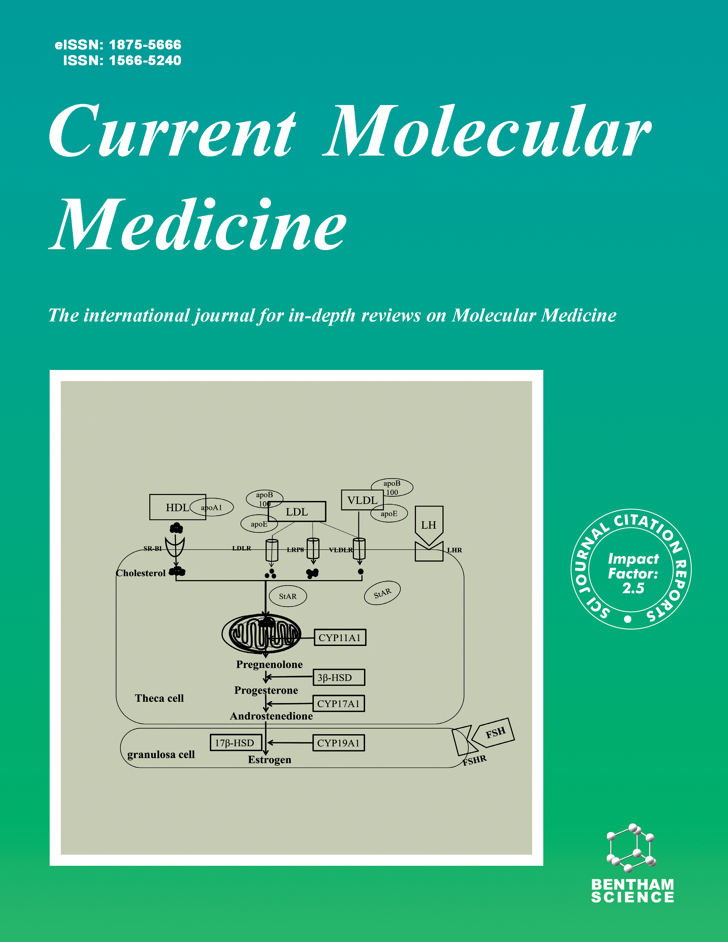
Current Molecular Medicine - Volume 18, Issue 5, 2018
Volume 18, Issue 5, 2018
-
-
VEGFR2 and VEGF-C Suppresses the Epithelial-Mesenchymal Transition Via YAP in Retinal Pigment Epithelial Cells
More LessBackground: Whereas retinal pigment epithelial (RPE) cells are known to secrete VEGF-A and VEGFR2, the functions of the autocrine VEGF signaling remain unclear. Meanwhile, anti-VEGF therapies have been applied routinely to treat ocular vascular diseases. Objective: The aim of this study was to determine the functions of the VEGF signaling in RPE cells and evaluate the consequences of its interruption. Methods: The genes involved in the VEGF and Hippo signal pathways were knocked down with siRNAs in both ARPE-19 cell line and human primary RPE cells via transient transfection whereas overexpression of VEGFR2 was mediated via adenovirus transduction. Expression of the epithelial-mesenchymal transition (EMT) markers and the downstream genes of YAP were determined by real-time PCR and Western Blot analysis. Immunofluorescence staining was utilized to determine gene expression in tissue and mouse samples. Results: Knockdown of VEGFR2 results in epithelial-mesenchymal transition in vitro and in vivo. Overexpression of VEGFR2 suppresses TGF β-mediated EMT in RPE cells. Loss of VEGF-C rather than VEGF-A induces EMT. Mechanistically, the VEGFR2 ablation-induced EMT in RPE cells is mediated by activation of YAP, an effector of the Hippo pathway. Finally, the immunohistochemical analysis of VEGFR2 and YAP in human proliferative vitreoretinopathy (PVR) membranes indicates a tendency of an inverse correlation between VEGFR2-positive and YAP-positive cells. Conclusions: Our results disclose unexpected novel roles of VEGFR2 and VEGF-C in the process of EMT of RPE cells and in the Hippo pathway. The data shown here demonstrated that VEGFR2 and VEGF-C are important to maintain the normal physiological state of RPE cells.
-
-
-
Mutation Analysis of Pre-mRNA Splicing Genes PRPF31, PRPF8, and SNRNP200 in Chinese Families with Autosomal Dominant Retinitis Pigmentosa
More LessBackground: To screen variants in pre-mRNA Splicing genes in 95 Chinese autosomal dominant retinitis pigmentosa (adRP) families. Methods: Clinical examination and pedigree analysis were performed. Targeted exome sequencing (TES) and / or Sanger sequencing were performed to detect the variants in genes of Splicing factors and conduct intra-familiar segregation analysis with DNA available. In silico analysis was performed to predict pathogenicity of variants in protein level and in vitro splicing assays were performed to compare splicing variants with their corresponding wildtype about their splicing effect. Results: In this study, total nine different variants were identified in PRPF31, SNRNP200, and PRPF8 respectively, including six PRPF31 variants [five novel variants 322+1G>A, c.527+2T>G, c.590T>C(p.Leu197Pro), c.1035_1036insGC (p.Pro346Argfs X18), and c.1224dupG (p.Gln409AlafsX66) plus one reported variant c.1060C>T (p.Arg354X)], a recurrent PRPF8 variant c.6930G>T (p.Arg2310Ser), two SNRNP200 variants [one heterozygous and homozygous SNRNP200 recurrent variant c.3260G>A (p.Ser1087Leu), and a reported heterozygous c.2042G>A(p.Arg681His)]. In family 20009, incomplete penetrance was observed. A novel PRPF31 missense variant c.590T>C (p.Leu197Pro) was predicted to be pathogenic in protein level via in silico analysis and in vitro splicing assay demonstrated that two novel splicing PRPF31 variants c.322+1G>A and c.527+2T>G affect splicing compared with the wildtype. Conclusions: In our studies, RP-causing variants of pre-mRNA Splicing genes (PRPF31, PRPF8 and SNRNP200) were identified in nine of the ninety-five adRP families respectively, which extend the spectra of RP variant and phenotype. And we provide the first example that SNRNP200-related RP can be caused by both heterozygous and homozygous variants of this gene.
-
-
-
Genetic Modifiers of Fetal Haemoglobin (HbF) and Phenotypic Severity in β-Thalassemia Patients
More LessAuthors: S.A.A. Razak, N.A.A. Murad, F. Masra, D.L.S. Chong, N. Abdullah, N. Jalil, H. Alauddin, R.Z.A.R. Sabudin, A. Ithnin, L.C. Khai, N.A. Aziz, Z. Muda, H. Ibrahim and Z.A. LatiffBackground: The phenotypic severity of β-thalassemia is highly modulated by three genetic modifiers: β-globin (HBB) mutations, co-inheritance of α-thalassemia and polymorphisms in the genes associated with fetal haemoglobin (HbF) production. This study was aimed to evaluate the effect of HbF related polymorphisms mainly in the HBB cluster, BCL11A (B-cell CLL/lymphoma 11A) and HBS1L-MYB (HBS1-like translational GTPase-MYB protooncogene, transcription factor) with regards to clinical severity. Methods: A total of 149 patients were included in the study. HBA and HBB mutations were characterised using multiplex PCR, Sanger sequencing and multiplex ligationdependent probe amplification. In addition, 35 HbF polymorphisms were genotyped using mass spectrometry and PCR-restriction fragment length polymorphism (PCRRFLP). The genotype-phenotype association was analysed using SPSS version 22. Results: Twenty-one HBB mutations were identified in the study population. Patients with HBB mutations had heterogeneous phenotypic severity due to the presence of other secondary modifiers. Co-inheritance of α-thalassemia (n = 12) alleviated disease severity of β-thalassemia. In addition, three polymorphisms (HBS1LMYB, rs4895441 [P = 0.008, odds ratio (OR) = 0.38 (0.18, 0.78)], rs9376092 [P = 0.030, OR = 0.36 (0.14, 0.90)]; and olfactory receptor [OR51B2] rs6578605 [P = 0.018, OR = 0.52 (0.31, 0.89)]) were associated with phenotypic severity. Secondary analysis of the association between single-nucleotide polymorphisms with HbF levels revealed three nominally significant SNPs: rs6934903, rs9376095 and rs9494149 in HBS1L-MYB. Conclusion: This study revealed 3 types of HbF polymorphisms that play an important role in ameliorating disease severity of β-thalassemia patients which may be useful as a predictive marker in clinical management.
-
-
-
Co-Existence of Novel PDE6A Mutations and A Recurrent RPGR Mutation: A Potential Explanation for Phenotypic Diversity in Female RPGR Mutation Carriers
More LessBackground: To report the co-existence of novel biallelic PDE6A mutations and heterozygous RPGR mutation in a Chinese female patient with retinitis pigmentosa (RP), and to analyze the intrafamilial phenotypic diversity. Methods: Three patients with retinopathy and four healthy family members were included in genetic and clinical analyses. Personal medical records were obtained from another four unaffected female family members who refused blood donation. Family history was carefully recorded. Each patient received comprehensive ophthalmic tests. Targeted next-generation sequencing (NGS) approach was performed on the proband to determine the retinopathy causative mutation for this family. In silico analysis was also applied to analyze the pathogenesis of identified mutations. Results: The two recruited male patients were diagnosed with RP, and the female patient RP sine pigmento (RPSP). Genetic assessments revealed a recurrent RPGR mutation, c.1926_1927insA, carried by all three patients and segregated the disease status. Three other unaffected female family members were confirmed as carriers for the identified RPGR mutation, and another four as obligate carriers. Interestingly, of all the eight female RPGR mutation carriers in this family, only one female developed retinal dystrophy. Comprehensive genetic analysis of this patient unraveled additional biallelic PDE6A mutations, c.[1066-9delT];[2324delG], carried solely by this individual. Conclusions: Taken together, we hypothesize that the phenotypic variability presented by female RPGR mutation carriers may be attributed to the co-existence of other disease-causative mutations. Our study also emphasizes the importance of comprehensive genetic analysis in these female carriers, which will contribute to better diagnosis, prognosis, and treatment for these patients.
-
-
-
MiR-19b Functions as a Potential Protector in Experimental Autoimmune Encephalomyelitis
More LessBackground: MicroRNA-19b (miR-19b) is essential in determining oligodendroglia proliferation. Phosphatase and tensin homologue on chromosome 10 (PTEN) is considered the target of miR-19b and participates in oligodendrocyte differentiation and proliferation. Methods: Murine EAE was induced by myelin oligodendrocyte glycoprotein (MOG35– 55). For EAE reversal, artificially synthesized agomiR-19b was intravenous injected after immunization. Results: We found that the expression of miR-19b is significantly reduced in an experimental autoimmune encephalomyelitis (EAE) mouse model. This downregulation, which is associated with the neurological scores, can be dramatically ameliorated by agomiR-19b. Our results show that agomiR-19b increases the expression of myelin basic protein (MBP) and cyclic nucleotide phosphodiesterase (CNP), which are regularly utilized as molecular markers of oligodendrocytes. Furthermore, our study also revealed that miR-19b probably affects the expression of PTEN in the EAE model. Conclusion: These results indicate that the restoration of miR-19b probably exerts its therapeutic effect by affecting PTEN in the pathogenesis of EAE.
-
-
-
Excessive Oxidative Stress in the Synergistic Effects of Shikonin on the Hyperthermia-Induced Apoptosis
More LessAuthors: J.-L. Piao, Y.-J. Jin, M.-L. Li, S.A. Zakki, L. Sun, Q.-W. Feng, D. Zhou, T. Kondo, Z.-G. Cui and H. InaderaBackground: Hyperthermia (HT) has been used widely for cancer therapy, and the development of modern devices has made it more efficient. Shikonin (SHK) is a natural naphthoquinone derivative from a Chinese herb. Although the anticancer properties of SHK are evident, the underlying molecular mechanisms are not fully understood. Objective: In this study, the effects of combining low doses of SHK with mild HT were investigated in the U937 cell line. Methods: The cells were subjected to HT at 44°C for 10 min with or without SHK pretreatment, and parameters reflecting apoptosis, ROS generation and intracellular calcium elevation were evaluated by using DNA fragmentation, flow cytometry, and western blot analyses. Results: SHK 0.5 μM significantly enhanced HT-induced apoptosis as indicated by DNA fragmentation and caspase-3 activation with increased generation of ROS and elevation of intracellular calcium. The combined treatment also synergistically activated proapoptotic proteins and inactivated anti-apoptotic proteins. Furthermore, the phosphorylation of JNK and PKC- δ and the dephosphorylation of ERK and AKT were the upstream effects that may have compounded the induction of apoptosis. The modulatory effects of HT and SHK were abrogated with the employment of NAC and JNK-IN-8 by inactivating the MAPK pathway and cleavage of caspase-3. Intracellular calcium was also elevated and was found to be responsible for the induction of cell death evident by the DNA fragmentation with or without the employment of BAPTA-AM. Conclusion: Conclusively, this study provides persuasive evidence that SHK in combination with HT is a propitious therapeutic way for augmentation of apoptosis and hence suggest a novel strategy for treating cancers.
-
-
-
Probable Mechanisms Involved in Immunotoxin Mediated Capillary Leak Syndrome (CLS) and Recently Developed Countering Strategies
More LessAuthors: B. Darvishi, L. Farahmand, N. Jalili and K. Majidzadeh-AAntibody-toxin fused agents or immunotoxins, are a newly engineered class of cytotoxic agents consisting of a bacterial or plant toxin moiety hooked up either to a monoclonal antibody or a specific growth factor. Nevertheless, acquiring a full potency in clinic is mostly restricted due to the Capillary leak syndrome (CLS), a serious immune provoked, life-threatening side effect, subsequent to the endothelial damage, resulting in fluid escape from the bloodstream into tissues including lungs, muscle and brain, developing organ failure and eventually death. Proposed underlying mechanisms include direct damage to endothelial cells, acute inflammation, Lymphokine-activated killer (LAK) cells engagement, alteration in cell-cell/cell-matrix connectivities and cytoskeletal dysfunction. Very poor biodistribution and heterogeneous extravasation pattern in tumor site result in accumulation of ITs close to the extravasation site, gradual toxin release and initiation of nearby endothelial cells lysis, secretion of pro-inflammatory cytokines, development of acute inflammation and engagement of Lymphokine-activated killer (LAK) cells. Intrinsic immunogenicity of applied toxin moiety is another important determinant of CLS incidence. Toxins with more intrinsic immunogenicity possess more probability for CLS development. Recently, development of new generations of antibodies and mutated toxins with conserved cytotoxicity has partly tapered risk of CLS development. Here, we describe probable mechanisms involved in CLS and introduce some of the recently applied strategies for lessening incidence of CLS as much as possible.
-
Volumes & issues
-
Volume 25 (2025)
-
Volume 24 (2024)
-
Volume 23 (2023)
-
Volume 22 (2022)
-
Volume 21 (2021)
-
Volume 20 (2020)
-
Volume 19 (2019)
-
Volume 18 (2018)
-
Volume 17 (2017)
-
Volume 16 (2016)
-
Volume 15 (2015)
-
Volume 14 (2014)
-
Volume 13 (2013)
-
Volume 12 (2012)
-
Volume 11 (2011)
-
Volume 10 (2010)
-
Volume 9 (2009)
-
Volume 8 (2008)
-
Volume 7 (2007)
-
Volume 6 (2006)
-
Volume 5 (2005)
-
Volume 4 (2004)
-
Volume 3 (2003)
-
Volume 2 (2002)
-
Volume 1 (2001)
Most Read This Month


