Planum Temporale Analysis Via a New Volumetric Method in Autoptic Brains of Demented and Psychotic Patients

- Authors: Petr Zach1, Zdena Kritofiková2, Jana Mrzílková3, Emerich Majer4, Pavel Selinger5, Filip Španiel6, Daniela Řípová7, Martina Kutová8, Jana Kenney9
-
View Affiliations Hide Affiliations1 Prague Psychiatric Center, Czech Republic, 2 Prague Psychiatric Center, Czech Republic, 3 Institute of Anatomy, Third Faculty of Medicine, Charles University, Czech Republic 4 Psychiatric Hospital Bohnice, Czech Republic 5 Faculty Hospital Bulovka, Czech Republic, 5Czech Academy of Science, Czech Republic 6 Prague Psychiatric Center, Czech Republic 7 Prague Psychiatric Center, Czech Republic 8 Institute of Anatomy, Third Faculty of Medicine, Charles University, Czech Republic 9 Czech Academy of Science, Czech Republic
- Source: Advances in Alzheimer's Research Volume 2 , pp 395-413
- Publication Date: September 2014
- Language: English
Planum Temporale Analysis Via a New Volumetric Method in Autoptic Brains of Demented and Psychotic Patients, Page 1 of 1
< Previous page | Next page > /docserver/preview/fulltext/9781608058525/chapter-14-1.gif
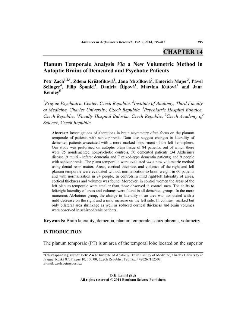
Investigations of alterations in brain asymmetry often focus on the planum temporale of patients with schizophrenia. Data also suggest changes in laterality of demented patients associated with a more marked impairment of the left hemisphere. Our study was performed on autoptic brain tissue of 84 patients, out of which there were 25 nondemented nonpsychotic controls, 50 demented patients (34 Alzheimer disease, 9 multi - infarct dementia and 7 mixed-type dementia patients) and 9 people with schizophrenia. The plana temporalia were evaluated via a new volumetric method using dental resin matter. Areas, cortical thickness and volumes of the right and left planum temporale were evaluated without normalization to brain weight in 60 patients and with normalization in 24 people. In controls, a mild right/left laterality of areas, cortical thickness and volumes was found. Moreover, in control women the areas of the left planum temporale were smaller than those observed in control men. The shifts to left/right laterality of areas and volumes were found in all demented groups. In the more numerous Alzheimer group, the change in laterality of an area was associated with a mild decrease on the right and a mild increase on the left side. In contrast, marked but only bilateral area shrinkage as well as reduced cortical thickness and brain volumes were observed in schizophrenic patients.
-
From This Site
/content/books/9781608058525.chapter-14dcterms_subject,pub_keyword-contentType:Journal -contentType:Figure -contentType:Table -contentType:SupplementaryData105

