oa Measurements of the Livers Total Blood Flow and Regional Perfusion

- Authors: Susanne Keiding1, Ole Lajord Munk2
-
View Affiliations Hide Affiliations1 PET Centre, Aarhus University Hospital, Aarhus, Denmark 2 PET Centre, Aarhus University Hospital, Aarhus, Denmark
- Source: Functional Molecular Imaging In Hepatology , pp 14-23
- Publication Date: May 2012
- Language: English
Measurements of the Livers Total Blood Flow and Regional Perfusion, Page 1 of 1
< Previous page | Next page > /docserver/preview/fulltext/9781608052905/chapter-2-1.gif
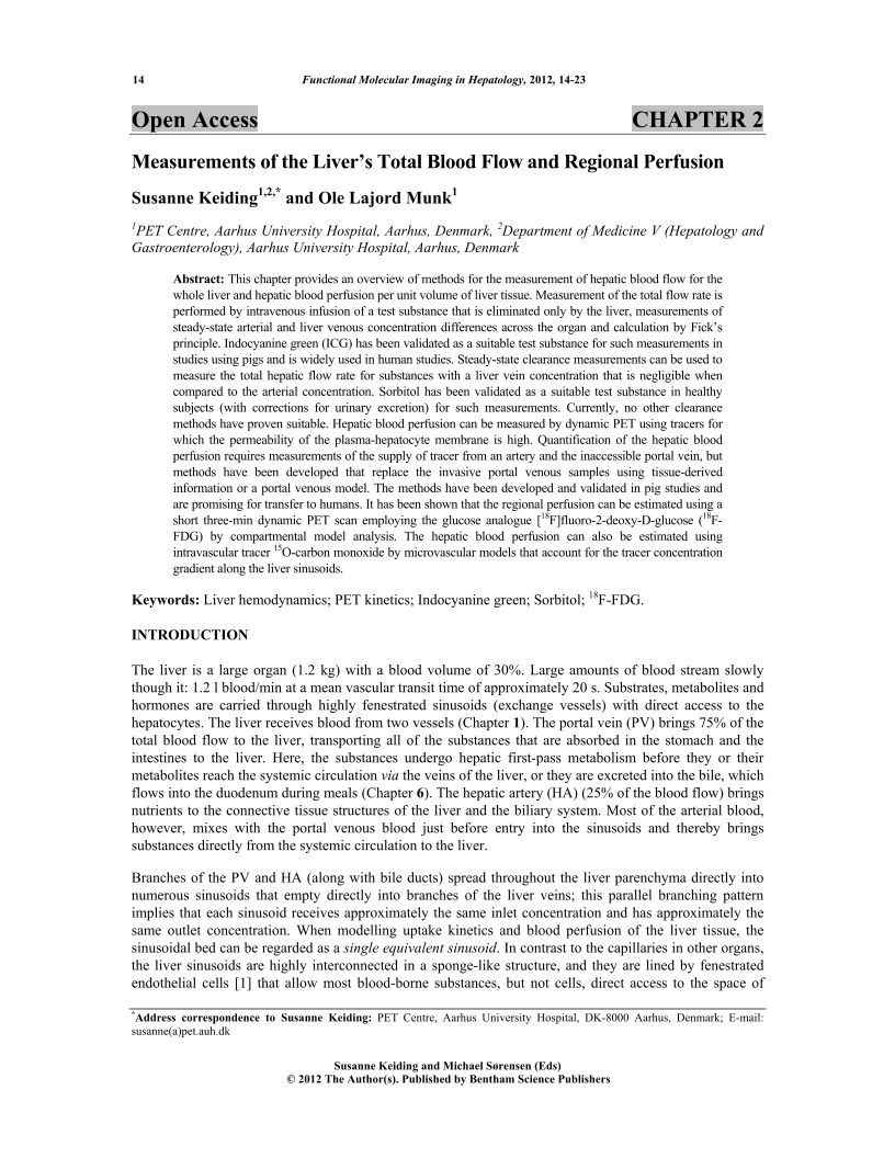
This chapter provides an overview of methods for the measurement of hepatic blood flow for the whole liver and hepatic blood perfusion per unit volume of liver tissue. Measurement of the total flow rate is performed by intravenous infusion of a test substance that is eliminated only by the liver, measurements of steady-state arterial and liver venous concentration differences across the organ and calculation by Ficks principle. Indocyanine green (ICG) has been validated as a suitable test substance for such measurements in studies using pigs and is widely used in human studies. Steady-state clearance measurements can be used to measure the total hepatic flow rate for substances with a liver vein concentration that is negligible when compared to the arterial concentration. Sorbitol has been validated as a suitable test substance in healthy subjects (with corrections for urinary excretion) for such measurements. Currently, no other clearance methods have proven suitable. Hepatic blood perfusion can be measured by dynamic PET using tracers for which the permeability of the plasma-hepatocyte membrane is high. Quantification of the hepatic blood perfusion requires measurements of the supply of tracer from an artery and the inaccessible portal vein, but methods have been developed that replace the invasive portal venous samples using tissue-derived information or a portal venous model. The methods have been developed and validated in pig studies and are promising for transfer to humans. It has been shown that the regional perfusion can be estimated using a short three-min dynamic PET scan employing the glucose analogue [18F]fluoro-2-deoxy-D-glucose (18FFDG) by compartmental model analysis. The hepatic blood perfusion can also be estimated using intravascular tracer 15O-carbon monoxide by microvascular models that account for the tracer concentration gradient along the liver sinusoids.
-
From This Site
/content/books/9781608052905.chapter-2dcterms_subject,pub_keyword-contentType:Journal -contentType:Figure -contentType:Table -contentType:SupplementaryData105

