Mistletoe Lectins, Structure and Function

- Authors: Wolfgang Voelter, Roland Wacker, Stanka Stoeva, Rania Tsitsilonis, Christian Betzel5
-
View Affiliations Hide Affiliations5 Abteilung fur Physikalische Biochemie des Physiologisch chemischen Instituts der Universitat Tubingen, Hoppe-Seyler-Str. 4, 72076 Tubingen, Germany
- Source: Frontiers in Natural Product Chemistry: Volume 1 , pp 149-162
- Publication Date: January 2005
- Language: English
Mistletoe Lectins, Structure and Function, Page 1 of 1
< Previous page | Next page > /docserver/preview/fulltext/9781608052127/chapter-16-1.gif
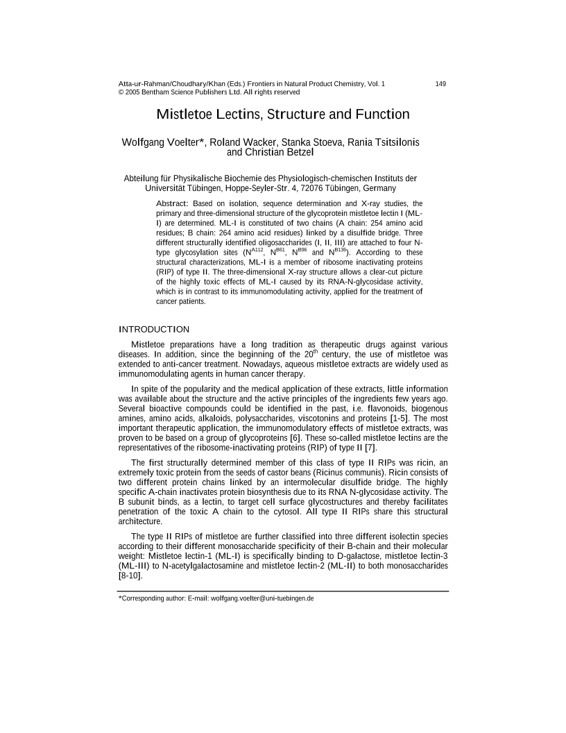
Based on isolation, sequence determination and X-ray studies, the primary and three-dimensional structure of the glycoprotein mistletoe lectin I (MLI) are determined. ML-I is constituted of two chains (A chain: 254 amino acid residues; B chain: 264 amino acid residues) linked by a disulfide bridge. Three different structurally identified oligosaccharides (I, II, III) are attached to four Ntype glycosylation sites (NA112, NB61, NB96 and NB136). According to these structural characterizations, ML-I is a member of ribosome inactivating proteins (RIP) of type II. The three-dimensional X-ray structure allows a clear-cut picture of the highly toxic effects of ML-I caused by its RNA-N-glycosidase activity, which is in contrast to its immunomodulating activity, applied for the treatment of cancer patients.
-
From This Site
/content/books/9781608052127.chapter-16dcterms_subject,pub_keyword-contentType:Journal -contentType:Figure -contentType:Table -contentType:SupplementaryData105

