Clinical Applications of in vivo Confocal Microscopy

- Authors: Antoine Labbé1, Bénédicte Dupas, Christophe Baudouin3
-
View Affiliations Hide Affiliations1 Department of Ophthalmology, Quinze-Vingts National Ophthalmology Hospital Paris & Vision Institute, INSERM UMRS 592, Pierre et Marie Curie-Paris 6 University, Paris 3 Department of Ophthalmology, Quinze-Vingts National Ophthalmology Hospital Paris & Vision Institute, INSERM UMRS 592, Pierre et Marie Curie-Paris 6 University, Paris
- Source: Diagnostic Technologies in Ophthalmology , pp 31-49
- Publication Date: May 2012
- Language: English
Clinical Applications of in vivo Confocal Microscopy, Page 1 of 1
< Previous page | Next page > /docserver/preview/fulltext/9781608051335/chapter-3-1.gif
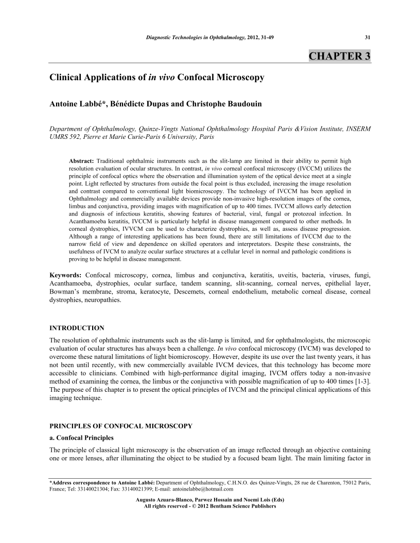
Traditional ophthalmic instruments such as the slit-lamp are limited in their ability to permit high resolution evaluation of ocular structures. In contrast, in vivo corneal confocal microscopy (IVCCM) utilizes the principle of confocal optics where the observation and illumination system of the optical device meet at a single point. Light reflected by structures from outside the focal point is thus excluded, increasing the image resolution and contrast compared to conventional light biomicroscopy. The technology of IVCCM has been applied in Ophthalmology and commercially available devices provide non-invasive high-resolution images of the cornea, limbus and conjunctiva, providing images with magnification of up to 400 times. IVCCM allows early detection and diagnosis of infectious keratitis, showing features of bacterial, viral, fungal or protozoal infection. In Acanthamoeba keratitis, IVCCM is particularly helpful in disease management compared to other methods. In corneal dystrophies, IVVCM can be used to characterize dystrophies, as well as, assess disease progression. Although a range of interesting applications has been found, there are still limitations of IVCCM due to the narrow field of view and dependence on skilled operators and interpretators. Despite these constraints, the usefulness of IVCM to analyze ocular surface structures at a cellular level in normal and pathologic conditions is proving to be helpful in disease management.
-
From This Site
/content/books/9781608051335.chapter-3dcterms_subject,pub_keyword-contentType:Journal -contentType:Figure -contentType:Table -contentType:SupplementaryData105

