Fetal 3D/4D Cardiovascular Imaging

- Authors: Toshiyuki Hata1, Junko Noguchi2
-
View Affiliations Hide Affiliations1 Department of Perinatology and Gynecology, Kagawa University, School of Medicine, 1750 1 Ikenobe, Miki Kagawa 761-0793, Japan. 2 Department of Nursing, Kagawa Prefectural College of Health Sciences, 281-1Murecho-hara, Takamatsu, Kagawa 761-0123, Japan
- Source: Current Topics on Fetal 3D/4D Ultrasound , pp 133-176
- Publication Date: March 2009
- Language: English
Fetal 3D/4D Cardiovascular Imaging, Page 1 of 1
< Previous page | Next page > /docserver/preview/fulltext/9781608050192/chapter-4-1.gif
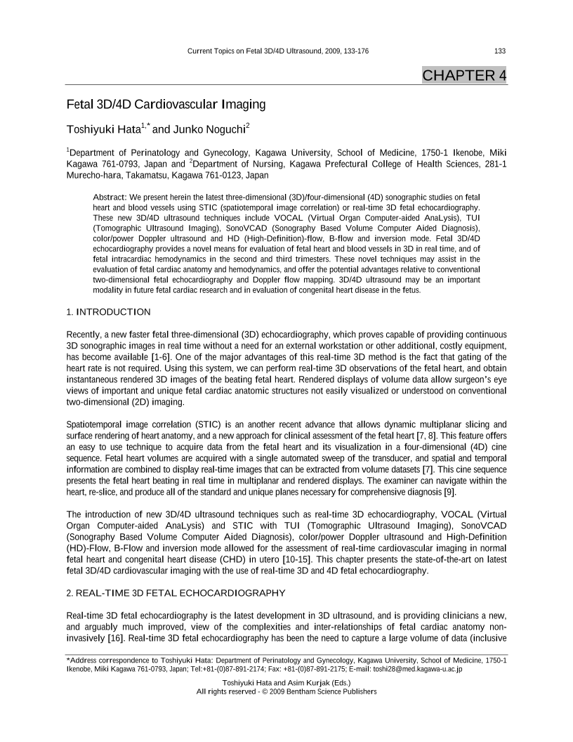
We present herein the latest three-dimensional (3D)/four-dimensional (4D) sonographic studies on fetal heart and blood vessels using STIC (spatiotemporal image correlation) or real-time 3D fetal echocardiography. These new 3D/4D ultrasound techniques include VOCAL (Virtual Organ Computer-aided AnaLysis), TUI (Tomographic Ultrasound Imaging), SonoVCAD (Sonography Based Volume Computer Aided Diagnosis), color/power Doppler ultrasound and HD (High-Definition)-flow, B-flow and inversion mode. Fetal 3D/4D echocardiography provides a novel means for evaluation of fetal heart and blood vessels in 3D in real time, and of fetal intracardiac hemodynamics in the second and third trimesters. These novel techniques may assist in the evaluation of fetal cardiac anatomy and hemodynamics, and offer the potential advantages relative to conventional two-dimensional fetal echocardiography and Doppler flow mapping. 3D/4D ultrasound may be an important modality in future fetal cardiac research and in evaluation of congenital heart disease in the fetus.
-
From This Site
/content/books/9781608050192.chapter-4dcterms_subject,pub_keyword-contentType:Journal -contentType:Figure -contentType:Table -contentType:SupplementaryData105

